Electrical Activity of the Heart
Electrical Activity of the Heart

Types of Cells in the Heart
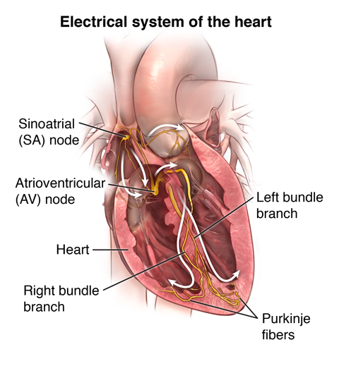
The heart is composed of two primary types of cells: 1. Pacemaker Cells (SA and AV Nodes): These cells are responsible for initiating and regulating the heartbeat. 2. Muscle Cells & His-Purkinje System (HPS): These cells are responsible for conducting the electrical signals throughout the heart to ensure coordinated contractions.
Pacemaker cells are found in key areas, specifically the Sinoatrial (SA) Node and the Atrioventricular (AV) Node.

SA Node vs. AV Node
SA Node: - The primary pacemaker of the heart. - Responsible for setting the pace of the heartbeat by generating electrical impulses.
AV Node: - Acts as a secondary pacemaker. - Slows down the electrical signal before it enters the ventricles, ensuring the atria have time to contract fully before the ventricles do.

Ion Movements and Membrane Potential
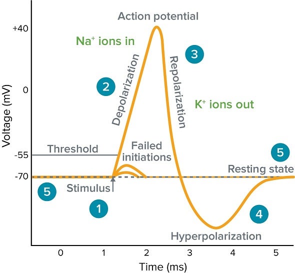
The electrical activity in the heart is heavily dependent on the movement of ions across the cell membranes. This movement changes the membrane potential and results in the generation of action potentials, crucial for the heart's function.
Key Ion Movements:
Calcium (Ca++) Influx in SA and AV Nodes:
- Ca++ ions moving inward is a significant event in the depolarization phase of pacemaker cells.
Potassium (K+) Efflux:
- K+ ions moving outward helps in repolarizing the membrane potential, bringing it back toward resting potential after a depolarization event.

Action Potential Phases in Pacemaker Cells
Phase 4 (Resting Potential):
- Membrane potential is gradually depolarized due to inward sodium (Na+) currents through the If (funny) channels.
Phase 0 (Depolarization Phase):
- Ca++ influx through L-type calcium channels (Ica.L) sharply depolarizes the cell.
Phase 3 (Repolarization Phase):
- K+ efflux through K+ channels (Iks and IKr) restores the cell to its resting membrane potential.

Membrane Potentials
The resting membrane potential of pacemaker cells is typically around -60mV to -40mV. During depolarization and repolarization, these potentials can swing:
- Depolarization: Membrane potential becomes more positive.
- Repolarization: Membrane potential returns to a more negative value.
These cyclic changes in membrane potential are what create the rhythmic heartbeat observed in a healthy heart.
Understanding these fundamentals of ion movement and membrane potentials is essential for comprehending the heart's electrical activity and its regulation, ensuring a systematic and coordinated heartbeat.
Here's an AI song: