Signal Transduction
Signal Transduction
G-Protein Coupled 7-Transmembrane Receptors
Overview

G-protein coupled receptors (GPCRs) are a vast and diverse group of membrane-bound receptors that play critical roles in transducing extracellular signals into intracellular responses. They interact with various ligands, including light, odorants, hormones (TSH, LH, FSH), small molecules, proteins, pheromones, chemokines, interleukins, and other signaling molecules. This group is characterized by seven transmembrane domains.
Activation and Signaling Mechanism

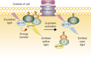
- Ligand Binding to Receptor: When a ligand binds to a GPCR, the receptor undergoes a conformational change.
- Activation of G-Protein: This change facilitates the exchange of GDP for GTP on the alpha subunit of the G-protein complex, activating the G-protein.
- GTPase Cycle:
- Inactive state: Gα bound to GDP.
- Active state: Gα bound to GTP and dissociates from the βγ complex.
- Activation of downstream effectors: Activated Gα and free βγ subunits interact with target proteins to propagate the signal.
G-Protein Activation of Adenylyl Cyclase

- Activated Gα subunit (Gs) stimulates adenylyl cyclase.
- Adenylyl Cyclase converts ATP to cAMP.
- cAMP Activation of Protein Kinase A (PKA): cAMP binds to PKA, releasing its catalytic subunits.
Protein Kinase A Activation

- Activation of CREB: PKA phosphorylates cAMP response element-binding protein (CREB), which then stimulates gene transcription.
- Activation of Glycogen Metabolism: PKA phosphorylates and activates phosphorylase kinase, which then activates glycogen phosphorylase, leading to the breakdown of glycogen into glucose-1-phosphate.
Cholera Toxin and Pertussis Toxin Effects

- Cholera Toxin: ADP-ribosylates Gαs, causing it to remain in the active GTP-bound state, resulting in persistent activation of adenylyl cyclase and elevated cAMP levels.
- Pertussis Toxin: ADP-ribosylates Gαi, preventing its interaction with the receptor and thus its activation. This blocks inhibition of adenylyl cyclase, leading to increased cAMP levels.
Phospholipase C (PLC) Activation
Phospholipase C Activation Pathway: Gq proteins activate PLC, which cleaves phosphatidylinositol 4,5-bisphosphate (PIP2) into diacylglycerol (DAG) and inositol trisphosphate (IP3).
- IP3: Opens Ca2+ channels on the endoplasmic reticulum, releasing Ca2+ into the cytosol.
- DAG: Activates protein kinase C (PKC), which then phosphorylates various target proteins.
Hormones Utilizing Calcium or Phosphatidylinositide Pathways
- Hormones like vasopressin, acetylcholine, ADH, cholecystokinin, oxytocin, and others mediate their effects through calcium or phosphatidylinositide signaling.
Summary of G-Protein Linked Receptors
- Stimulatory and Inhibitory Pathways: GPCRs can either stimulate or inhibit adenylyl cyclase activity, thus increasing or decreasing cAMP levels in response to various ligands.
- Phospholipase C Pathways: Activation leads to the production of DAG and IP3, resulting in the activation of PKC and a rise in intracellular calcium levels.
Receptor Tyrosine Kinases (RTKs)

Key Features
- Structure: Single transmembrane-spanning proteins with an extracellular ligand-binding domain, a single transmembrane helix, and an intracellular tyrosine kinase domain.
- Activation: Ligand binding induces receptor dimerization and autophosphorylation on tyrosine residues within the intracellular domain.
- Downstream Signaling Pathways:
- Phospholipase C-γ (PLC-γ): Activated by tyrosine phosphorylation and hydrolyzes PIP2, similar to GPCR-activated PLC.
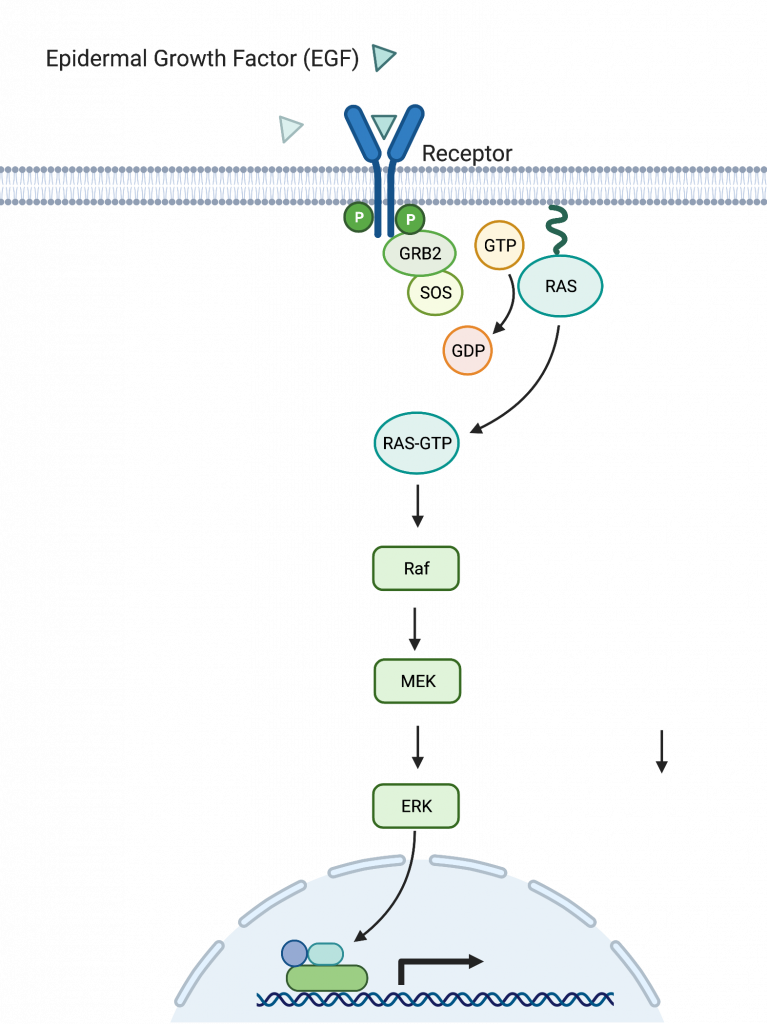
- Ras-Raf-MEK-ERK Pathway: Involves a cascade of protein kinases leading to the phosphorylation and activation of various transcription factors.
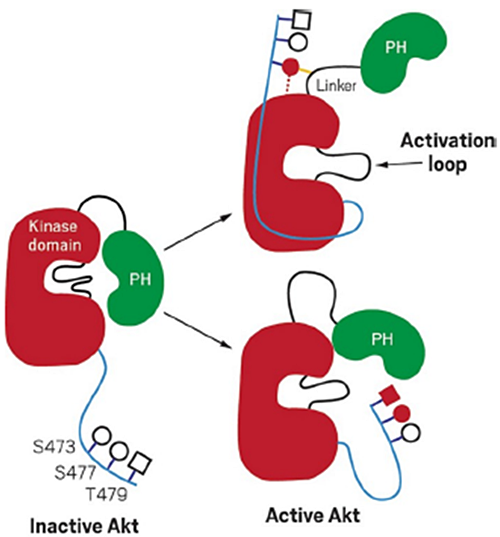
- PI3-Kinase Pathway: Produces PIP3, aiding in the activation of AKT (PKB), which regulates processes such as metabolism, survival, and growth.
JAK-STAT Pathway

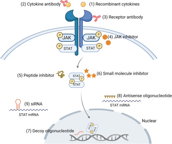
- Activation: Ligand binding to cytokine receptors results in the activation and dimerization of Janus kinases (JAKs).
- Signal Transduction: Activated JAKs phosphorylate Signal Transducers and Activators of Transcription (STAT) proteins, which then translocate to the nucleus to modulate gene expression.
Termination of Signaling

- Phosphatases: PTEN and SHIP dephosphorylate PIP3 to PIP2, terminating PI3K-AKT signaling.
- Internalization: Insulin-receptor complexes are endocytosed and degraded to terminate the signal.