Local Blood Flow and Exercise
Local Blood Flow and Exercise
Learning ObjectivesBy the end of this session, you should be able to:
- Describe how local metabolic demand affects blood flow in a tissue
- Explain acute and long-term control of local blood flow
- List important vasoconstrictor and vasodilator factors
- Explain how exercise affects blood flow to the skeletal muscle, blood pressure, and cardiac output.

Importance of Local Blood Flow Control
- The blood flow to each tissue is always controlled at a level only slightly more than required to maintain full tissue oxygenation.
- Tissues almost never suffer from oxygen nutritional deficiency, and yet the workload on the heart is kept at a minimum.
- For example, in the resting state, the metabolic activity of the muscles is very low and so is the blood flow (4 ml/min/100 g muscle).
- During heavy exercise, muscle metabolic activity increases more than 60-fold and the blood flow as much as 20-fold (80 ml/min/100 g muscle).

Local and Humoral Control
Mainly dependent on oxygen availability:
- Acute Control: Local vasodilation/vasoconstriction within seconds to minutes.
- Long-term Control: Increase/decrease in the physical sizes and numbers of blood vessels over days, weeks, and months.
Acute Control of Local Blood Flow

1. Vasodilator Theory
- Decreased availability of oxygen leads to adenosine release followed by vasodilation.
- Best known in coronary arteries.
- Researchers believe adenosine is an important controller of blood flow in skeletal muscle and other tissues as well.
2. Oxygen Lack Theory
- Also known as nutrient lack theory.
- Local precapillary and metarteriole sphincters open and close cyclically several times per minute.
- The cyclic opening and closing requires oxygen (and nutrients).
- When the oxygen concentration is high, the sphincters stay closed until the tissue consumes the excess oxygen.
- When the oxygen concentration falls low enough, the sphincters open.
General Mechanisms of Local Blood Flow
- Present in almost all tissues.
- Special areas have distinctly different mechanisms:
- Kidneys: Tubuloglomerular feedback.
- Brain: Concentrations of carbon dioxide and hydrogen ions play very prominent roles. Increase in either or both dilates the cerebral blood vessels.
- When microvascular blood flow increases, upstream arteries are dilated through the endothelium-derived relaxing factor (EDRF, mostly nitric oxide).
Autoregulation of Blood Flow in Response to Acute BP Change
- When BP increases acutely, there is an immediate rise in local blood flow.
- Within less than a minute, the blood flow in most tissues returns to almost the normal level, even though the arterial pressure is kept elevated. This is called "autoregulation of blood flow."

1. Metabolic Theory
- A BP increase leads to an increase in blood flow, followed by excess oxygen/nutrients, which leads to vasoconstriction and a return to normal blood flow.
2. Myogenic Theory
- BP increase leads to increased blood flow, vessel stretch, stretch-induced depolarization, vasoconstriction, and normal blood flow.
- The precise mechanism behind stretch-induced depolarization is not completely understood but involves ion channels and extracellular proteins.
- The myogenic mechanism may be important in preventing excessive stretch of blood vessels.
Long-term Blood Flow Regulation
- Acute mechanisms for blood flow control are effective but cannot fully adjust to the requirements.
- Long-term regulation develops over hours, days, and weeks, giving far more complete regulation.
- The principal mechanism of long-term regulation is to change "tissue vascularity," involving the actual physical reconstruction of the tissue vasculature.
- Oxygen is important for both acute control and long-term control of blood flow.
- Growth factors like VEGF, fibroblast growth factor, and angiogenin are involved in increasing the growth of new vessels.
- Vascularity is determined by the maximal level of blood flow need rather than by average need.
Development of Collateral Circulation
- This is a phenomenon of long-term local blood flow regulation.
- When a blood vessel is blocked, a new vascular channel (collateral vessels) usually develops around the blockage and allows at least partial supply of blood.
- Initial dilation of small vascular loops that already connect the vessel above the blockage to the vessel below (within 1-2 minutes).
- Further dilation occurred within the ensuing hours, reaching ~ half of the needed flow over one day and often full capacity within a few days.
- Continued growth of collateral vessels, almost always forming many small vessels rather than one large vessel.

Humoral Control of the Circulation
- Humoral control means control by hormones and ions. Below are some of the important vasoconstrictor and vasodilator factors:
- Vasoconstrictor agents: Norepinephrine, Epinephrine (may also dilate), Angiotensin II, Vasopressin, Endothelin.
- Vasodilator agents: Bradykinin, Histamine, Potassium (K+), Magnesium (Mg2+), Hydrogen ions (H+), Carbon dioxide (CO2).

- Vasoconstrictor agents: Norepinephrine, Epinephrine (may also dilate), Angiotensin II, Vasopressin, Endothelin.
- Vasodilator agents: Bradykinin, Histamine, Potassium (K+), Magnesium (Mg2+), Hydrogen ions (H+), Carbon dioxide (CO2).
Muscle Blood Flow and Cardiac Output During Exercise
Blood Flow in Skeletal Muscle- During strenuous exercise, skeletal muscle requires large amounts of blood flow.
- Blood flow is not as high during contractions because the blood vessels are ‘squeezed’.

Mechanisms of Blood Flow Increase in Skeletal Muscle During Exercise
- Local blood flow increase in skeletal muscle:
- Oxygen is used up rapidly, resulting in the release of vasodilators (e.g., CO2, H+, Adenosine, Lactose, and K+).
- Loss of arteriolar wall contraction due to oxygen lack.
- Massive systemic sympathetic discharge leading to:
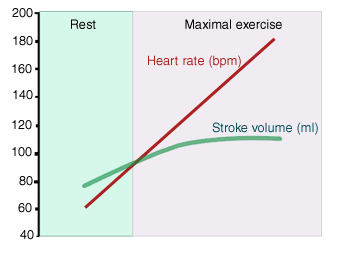
- Increased HR and contractility, causing a direct increase in cardiac output (CO).
- Systemic α1 vasoconstriction (and β2 vasodilation in skeletal muscle blood vessel) causing redirection of blood to skeletal muscle.
- Venoconstriction increasing mean systemic filling pressure/venous return, leading to an increase in CO.

Increase in Cardiac Output During Exercise
- Many different physiological effects occur to increase cardiac output (CO) approximately in proportion to the degree of exercise.
- The ability to increase CO for delivery of oxygen and nutrients to the muscles during exercise is crucial for continued muscle work.

Cardiac Function During Exercise
- The cardiac function curve shifts up due to increased HR and contractility from sympathetic stimulation.
- The venous return curve shifts right due to:
- Venoconstriction.
- Tensing of abdominal and other skeletal muscles.
- A decrease in TPR (total peripheral resistance) increases the slope of both curves, contributing to the increase in CO.
- At rest, cardiac function and venous return curves for normal circulation cross at point A, giving a CO of about 5 L/min.
- During heavy exercise, both cardiac function and venous return curves change significantly, yielding a CO of over 20 L/min (point B).