Heart Histology: Structure and Function of the Heart Wall
Heart Histology: Structure and Function of the Heart Wall
:max_bytes(150000):strip_icc()/the-heart-wall-4022792-FINAL-ff0aca97377c4fe9aeef72b044138011.png)
1. Heart Wall Layers
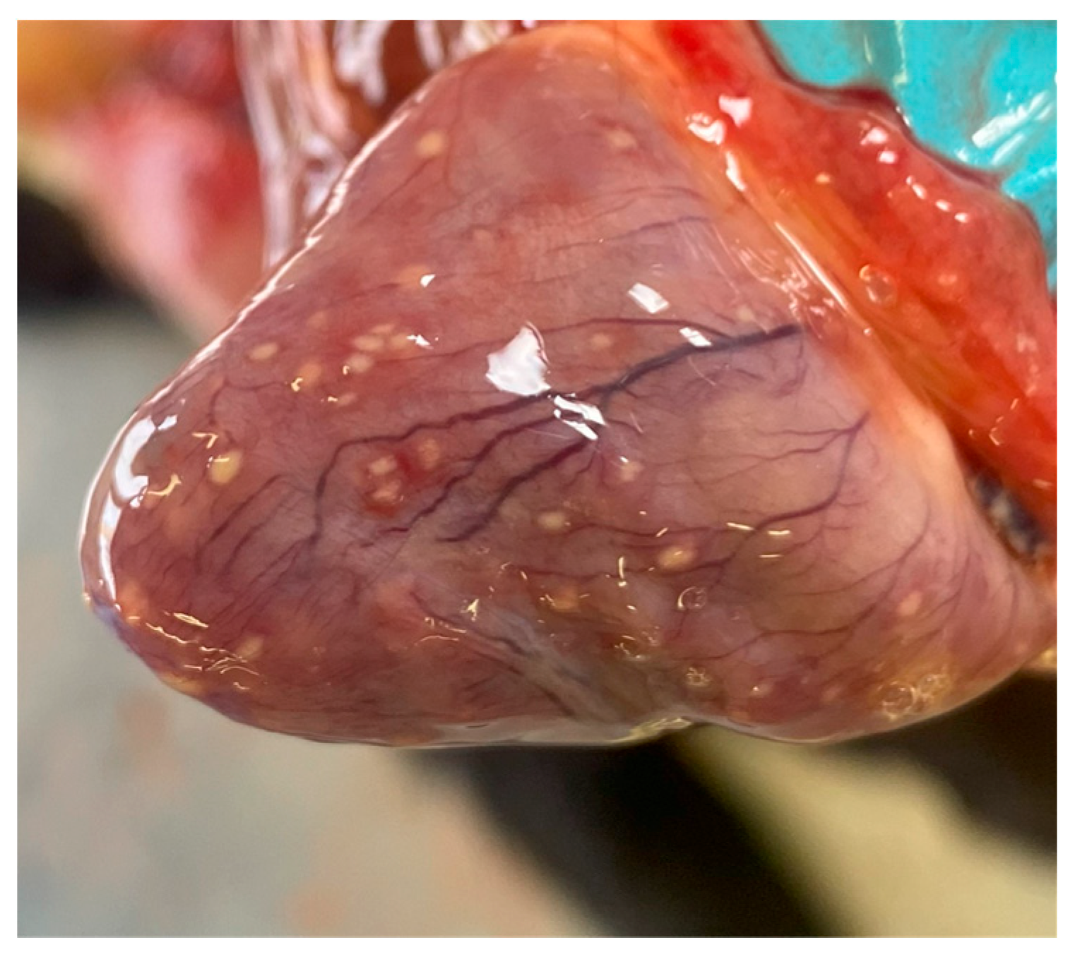
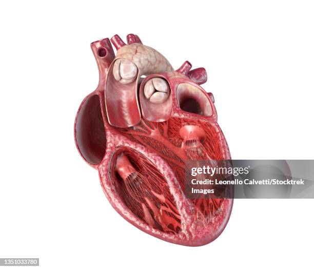
The heart wall consists of three distinct layers: the epicardium, myocardium, and endocardium, with the presence of visceral and parietal pericardium surrounding the heart.
Epicardium
- Composition: Mesothelium (simple squamous/cuboidal epithelium) + connective tissue (CT)
- Components: Adipose tissue, blood vessels, and parasympathetic ganglia (not always visible)
- Function: The mesothelium secretes serous fluid to reduce friction
Myocardium
- Composition: Cardiac muscle tissue + connective tissue
- Characteristics:
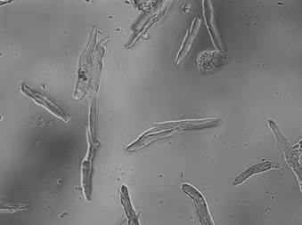
- Single central nucleus
- Branched myocytes
- Intercalated discs
- Striated myofibrils (sarcomeres)
- Absence of mitosis (no hyperplasia)

Endocardium
- Composition: Endothelium (simple squamous epithelium) + areolar connective tissue
- Components: Purkinje fibers (not always visible), blood inside the heart
2. Specialized Heart Cells
Atrial Myocytes- Endocrine Function: Production and secretion of atrial natriuretic peptide (ANP)
- Location: Principally in the atria
- Trigger for Secretion: Increased atrial pressure
- Function of ANP: Lowers blood pressure by promoting sodium excretion (natriuresis) and reducing blood volume, contrary to aldosterone
Purkinje Fibers
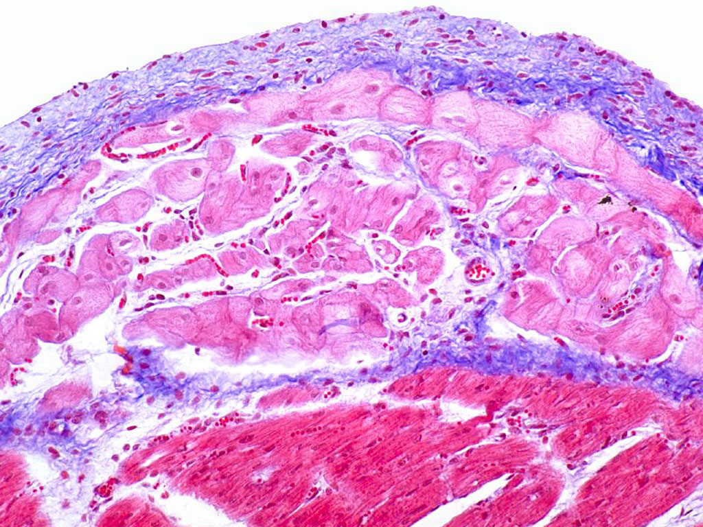
- Histological Differences from Regular Myocytes:
- Larger size
- Located in the subendocardium
- Glycogen-rich cytoplasm pushes myofibrils to the periphery
- Connected by desmosomes and gap junctions (no intercalated discs)
- Electrical Function: Rapid conduction of electrical impulses through the AV node, bundle branches, and into the ventricles
3. Heart Valves
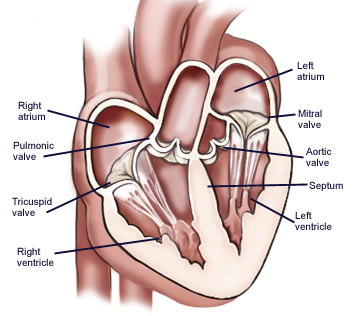
[h3]Structure- Layers: Lined by endocardium and have a core of fibrous connective tissue
- Components: Chordae tendineae and papillary muscles
Histological Composition
- AV Valves (Tricuspid and Mitral): Dense collagen core with endocardium lining
- Semilunar Valves (Aortic and Pulmonary): Similar structure to AV valves
- Clinical Relevance: Pathological conditions such as endocarditis, often linked to rheumatic fever (streptococcal infection), can cause valve dysfunction

4. Clinical Correlations
Pericarditis
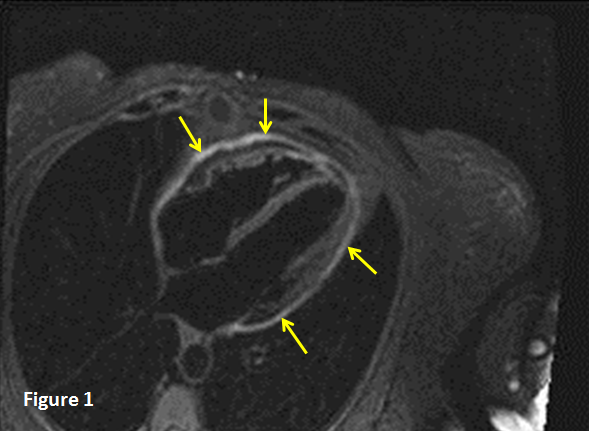
- Causes: Infection, autoimmune disorders, and other inflammations leading to a swollen pericardial cavity
- Diagnostic Imaging: MRI can reveal pericardial effusion and inflammation
- Pathology: Death of cardiac tissue due to ischemia, identified by the release of cardiac troponins
- Histology: Presence of granulation tissue, fibrosis, and disarrayed muscle cells
- Etiology: Infection (bacterial or fungal) or autoimmune response leading to inflammation of heart valves
- Histology: Inflammatory infiltrate, Anitschkow cells in rheumatic heart disease, and vegetations seen on echocardiograms
- Normal: Adaptive response to increased workload as seen in athletes
- Abnormal: Pathological in hypertrophic cardiomyopathy (HCM) and linked with genetic mutations leading to disorganized myocyte structure
- Myopathy: General muscle tissue disorder
- Myocarditis: Inflammation of the myocardium
- LVH (Left Ventricular Hypertrophy)[/strong]: Result of systemic hypertension, increased size of myofibers
 Great Vessels
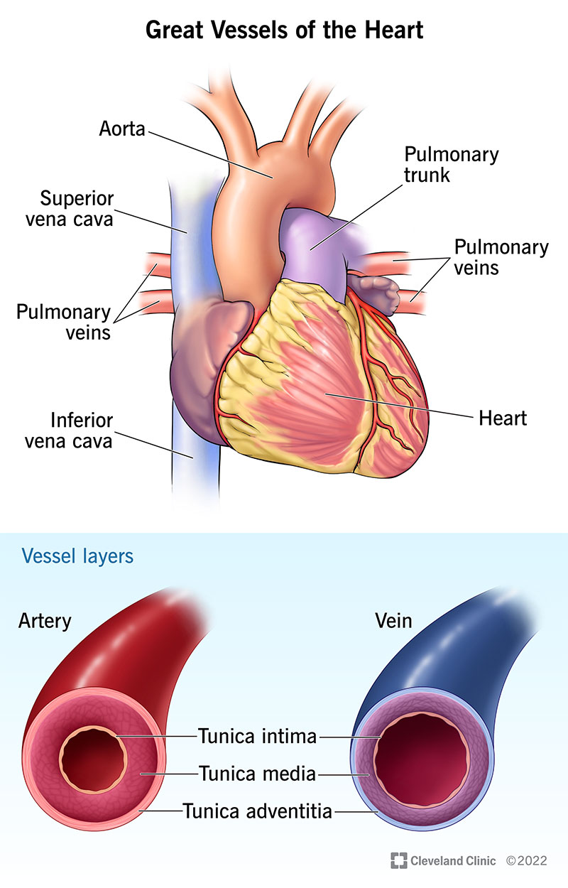
- Aorta: Thick tunica media with robust elastic lamina, prone to aneurysm in disorders like Marfan's syndrome
- Vena Cava: Prominent tunica adventitia with longitudinal muscle bundles
- Types: Continuous, fenestrated, and sinusoidal, each adapted to specific tissue needs
- Function: Facilitating exchange of gases, nutrients, and waste between blood and tissues