Superficial Anatomy of the Back
Overview of Back Musculature
The superficial muscles of the back are primarily responsible for shoulder and upper arm movements. These muscles include the trapezius, latissimus dorsi, levator scapulae, and rhomboid muscles. Each muscle has distinct origins, insertions, innervations, and functions, contributing to a wide range of upper body movements and stability. You need to know the muscle names, innervations, blood supply, and insertions & attachments. From high to low yield, it would be the names > actions > innervations > blood supply > insertions > attachments.
I spent way too much time on the first quiz memorizing highly esoteric functions when the actions and rudimentary details are truly the most important. I'd recommend getting out a whiteboard and writing down all the muscles that you need to know before the TBLs and charting all the details about each one. Use your own body as well to intuit how it all connects.
Trapezius Muscle
:max_bytes(150000):strip_icc()/GettyImages-147219941-56a05efe3df78cafdaa14c5f.jpg)
- Origin: The trapezius muscle originates from the external occipital protuberance, the nuchal ligament, and the spinous C7 to T12 vertebrae processes.
- Insertion: It inserts onto the lateral third of the clavicle, the acromion, and the spine of the scapula.
- Innervation: The muscle is innervated by the accessory nerve (cranial nerve XI) and the cervical spinal nerves (C3 and C4).
- Function: The trapezius muscle elevates, retracts, and rotates the scapula. It can also extend the neck.
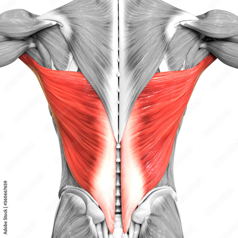
Latissimus Dorsi Muscle

- Origin: This muscle originates from the spinous processes of T7 to T12, the thoracolumbar fascia, the iliac crest, and the inferior three or four ribs.
- Insertion: It inserts into the intertubercular sulcus of the humerus.
- Innervation: The thoracodorsal nerve (C6, C7, and C8) innervates the latissimus dorsi.
- Function: The latissimus dorsi extends, adducts, and medially rotates the humerus. It also plays a role in movements such as pulling and lifting.
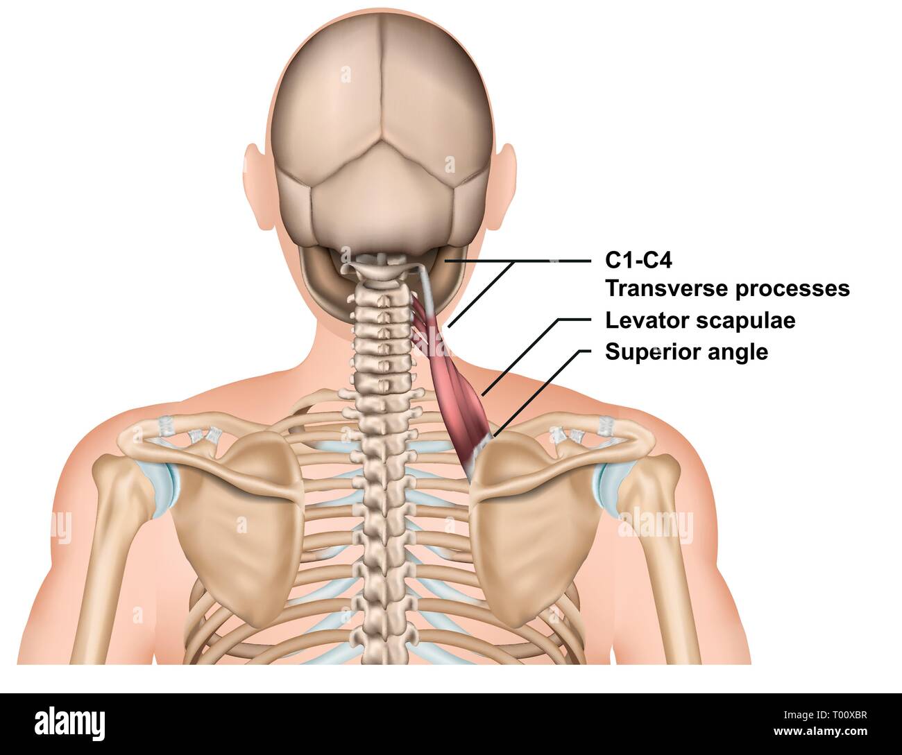
Levator Scapulae Muscle

- Origin: The levator scapulae originates from the transverse processes of C1 to C4 vertebrae.
- Insertion: It inserts onto the medial border of the scapula, between the superior angle and the spine.
- Innervation: The dorsal scapular nerve (C5) and cervical nerves (C3 and C4) provide innervation.
- Function: This muscle elevates the scapula and tilts the glenoid cavity inferiorly by rotating the scapula.
Rhomboid Major and Minor Muscles

- Origin: The rhomboid minor originates from the nuchal ligament and the spinous processes of C7 and T1. The rhomboid major originates from the spinous processes of T2 to T5.
- Insertion: Both muscles insert onto the medial border of the scapula. The rhomboid minor inserts at the level of the spine of the scapula, while the rhomboid major inserts below the spine.
- Innervation: The dorsal scapular nerve (C4 and C5) innervates both muscles.
- Function: The rhomboid muscles retract the scapula, pulling it towards the vertebral column, and rotate it to depress the glenoid cavity. They also help to fix the scapula to the thoracic wall.
Intrinsic Muscles: The intrinsic muscles, also known as the deep muscles of the back, are primarily involved in the movements and stabilization of the vertebral column. These include:
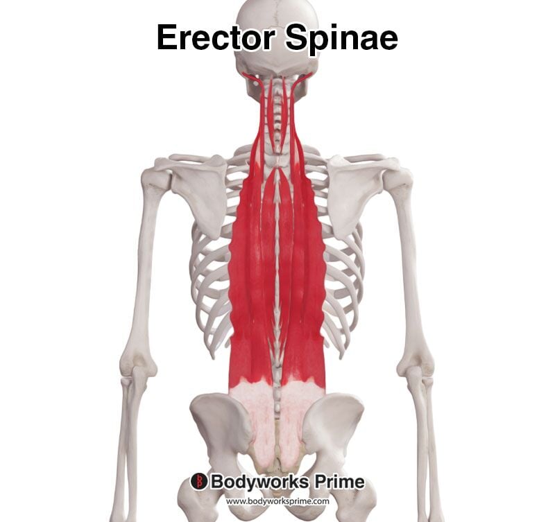
Erector Spinae:
Divisions: Iliocostalis, longissimus, and spinalis.
Action: Extend and laterally flex the vertebral column.
Innervation: Dorsal rami of spinal nerves.
I don't remember really having to know this muscle, except for the fact that it's deep.
Transversospinalis Group:

- Components: Semispinalis, multifidus, and rotatores muscles.
- Action: Rotate and extend the vertebral column.
- Innervation: Dorsal rami of spinal nerves.
- Again, it's sufficient just to know that they exist.
Surface Anatomy Landmarks
Key landmarks on the back are used in clinical practice to identify underlying structures and guide interventions. They love these:
- Vertebra Prominens (C7): The most prominent spinous process at the base of the neck.
:watermark(/images/watermark_only.png,0,0,0):watermark(/images/logo_url.png,-10,-10,0):format(jpeg)/images/anatomy_term/vertebra-prominens-2/5eNluhwCSK7Ue76NWAxBrw_Vertebra_Prominens_2.png)
- Scapular Spine: Runs horizontally across the upper back and aligns with the T3 vertebra.
- Inferior Angle of the Scapula: Located at the level of the T7 vertebra.
- Iliac Crest: The upper margin of the pelvis, aligned with the L4 vertebra.

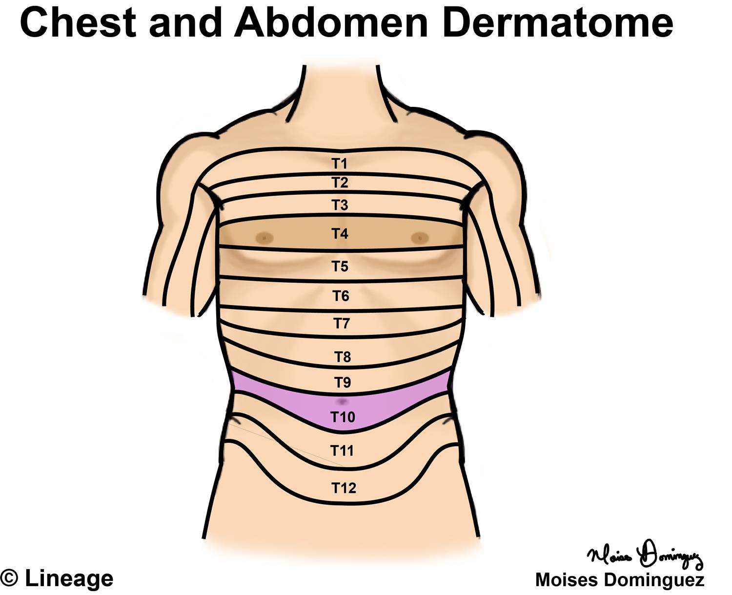
The most important landmarks are C7 (vertebra prominent, spinous process), T10 (umbilicus "belly buTEN"), T4 (nipple "Teat pore"),

* Note from an M2: You should know T10 and T4. They come up time and time again.
Cutaneous Innervation of the Back
The dorsal rami of the spinal nerves innervate the skin over the back. These nerves provide sensory innervation to the skin and deeper structures in a segmental pattern. Each spinal nerve innervates a specific dermatome, an area of skin supplied by a single spinal nerve. The cutaneous innervation is straight-forward.
- Cervical Region: Innervated by the dorsal rami of cervical spinal nerves.
- Thoracic Region: Innervated by the dorsal rami of thoracic spinal nerves.
- Lumbar Region: Innervated by the dorsal rami of lumbar spinal nerves.
- Sacral Region: Innervated by the dorsal rami of sacral spinal nerves.
Why does this matter?
Understanding the superficial anatomy of the back is crucial for clinical examinations and procedures. For instance:
- Physical Examination: Knowledge of the back muscles aids in diagnosing muscular or nerve injuries.
- Surgical Procedures: Accurate anatomical knowledge is essential during surgeries involving the back to avoid damaging vital structures.
- Injections and Lumbar Puncture: Familiarity with dermatomal innervation helps administer local anesthesia and perform lumbar punctures. Lumbar puncture questions come up a lot.
Study Song
Here's a fun study-break song that explains some of this content: