Middle Mediastinum, Pericardium, and Heart
Middle Mediastinum, Pericardium, and Heart
Note from an M2: Memorize the vascular structures on the heart. Remember "tri" before you "buy".
Important Anatomical Points:
- Inferior Border: Determines the lower limits of the lungs.
- Pleural Reflection: The line where the pleura changes direction.
- Bare Area of Pericardium: An area where the pericardium is exposed without pleural covering.
Topography of Lungs

Mediastinum
Boundaries and Subdivisions
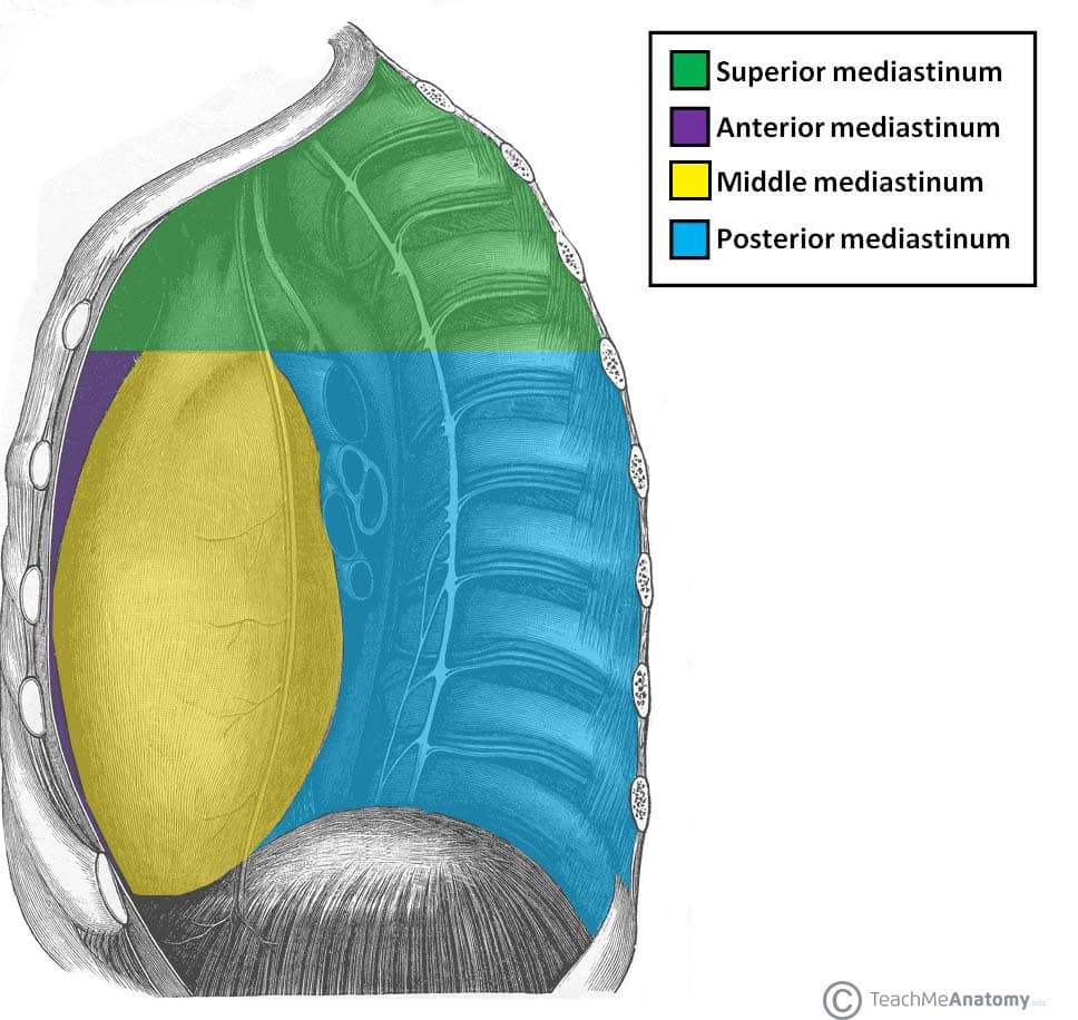
The mediastinum is centrally located in the thoracic cavity and is divided into four subdivisions:
- Superior Mediastinum: Located above the plane of the thoracic inlet
- Anterior Mediastinum: Situated in front of the pericardium
- Middle Mediastinum: Contains the heart and roots of the great vessels
- Posterior Mediastinum: Lies behind the pericardium

Important Contents in the Middle Mediastinum
- Pericardium: A fibrous sac encompassing the heart
- Heart
- Roots of the Great Vessels
Pericardial Sac and Pericardial Cavity
The pericardial sac comprises a fibrous outer layer and a serous inner layer lined with mesothelium. It encloses the pericardial cavity, a potential space filled with serous fluid.
Heart Anatomy
Positions and Surfaces:
- Base: Primarily the left atrium, lies opposite the apex
- Apex: The pointed end of the heart directed downward, forward, and to the left
- Sternocostal Surface: Anterior, facing the sternum and ribs
- Diaphragmatic Surface: Inferior, resting on the diaphragm
- Pulmonary Surfaces: Facing the lungs on either side

Sulci:
- Atrioventricular (Coronary) Sulcus: Separates the atria from the ventricles
- Interventricular Sulci: Anterior and posterior, marking the separation between the right and left ventricles
Heart Valves and Sounds
The heart's valves, including the mitral and tricuspid valves, ensure unidirectional blood flow. Auscultatory areas for heart valve sounds are located at specific intercostal spaces.

Coronary Circulation
Coronary arteries and veins supply and drain the myocardium. Notable coronary arteries include the right and left coronary arteries, with branches like the anterior interventricular and circumflex arteries. The pericardium is associated with clinical conditions such as pericarditis, pericardial effusion, and cardiac tamponade.
Conducting System of the Heart
The heart's conducting system includes the sinoatrial node, atrioventricular node, and Purkinje fibers, which coordinate heart contractions.

Clinical Correlations
Pathologies related to the heart include conditions like infective endocarditis, which often involves bacterial infection of the heart valves.

Pulmonic and Systemic Circuits
The heart is central to two main circuits: the pulmonic circuit, where the right ventricle pumps blood to the lungs, and the systemic circuit, where the left ventricle pumps blood to the body. The left ventricular wall is inherently thicker due to the higher resistance it must overcome.

Here is a song to practice: