Internal Genital Organs and Pelvic Vasculature
Internal Genital Organs and Pelvic Vasculature
Peritoneum
The peritoneum is a continuous serous membrane that lines the abdominal cavity and covers the abdominal organs. It plays a significant role in providing support and creating pathways for blood vessels, nerves, and lymphatics. In the context of the pelvis, the peritoneum extends from the abdomen to enter the true pelvis but does not come into contact with the pelvic diaphragm. 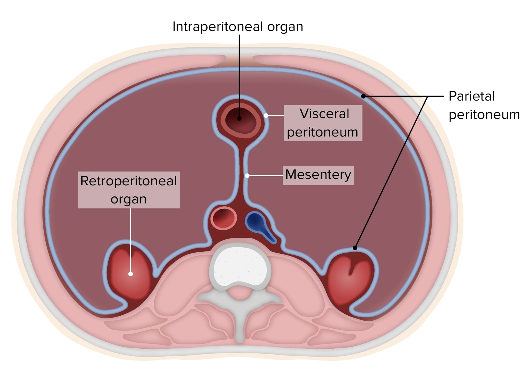
Male Peritoneum
In males, the peritoneum covers several pelvic structures:
- Urinary Bladder: As the bladder expands, the peritoneum on the superior surface can be lifted above the pubis into the abdominal cavity.
- Rectum: The rectum is subperitoneal at its inferior third, partially covered at its middle third, and forms the sigmoid mesocolon at its superior third.
- Rectovesical Pouch: A significant space formed by the peritoneum between the bladder and rectum.

Female Peritoneum
In females, the peritoneum covers additional reproductive organs:
- Urinary Bladder: Similar to males, it can be lifted above the pubis.

- Uterus and Fallopian Tubes: The peritoneum covers part of the uterus and all of the uterine tubes, broad ligaments, and ovaries.

- Rectouterine Pouch (Pouch of Douglas): This is located between the rectum and the uterus.

- Vesicouterine Pouch: This lies between the bladder and the uterus.
Rectum
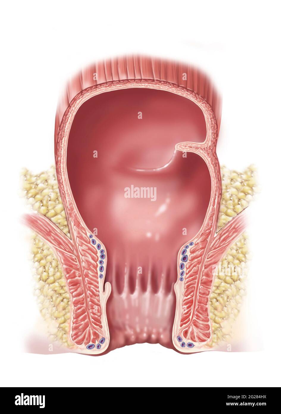
The rectum is the terminal portion of the large intestine, extending from the rectosigmoid junction at the level of the third sacral vertebra (S3) to the anus. It's important to note the following about its anatomical features:
- Transverse Rectal Folds: These folds help retain fecal matter.
- Rectal Ampulla: An anatomical dilation that stores feces before they are expelled.
- Anal Canal: This is the continuation of the rectal ampulla and terminates in the anus.
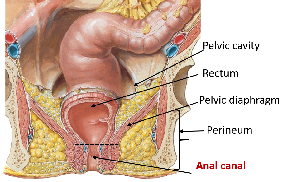
Peritoneal Coverage of the Rectum
- Inferior Third: Subperitoneal
- Middle Third: Anterior surface only
- Superior Third: Anterior and lateral sides

Male Internal Genital Organs
Prostate
The prostate gland surrounds the neck of the bladder and the prostatic urethra. Significant anatomical features include:
- Prostatic Utricle: A small indentation in the prostatic urethra.
- Ejaculatory Ducts: Tubes that pass through the prostate and open into the prostatic urethra.

- Prostatic Sinus: A groove at either side of the urethral crest where the prostatic ducts open.

Seminal Vesicles
These are tubular glands that secrete fluid that partly composes the semen. They are located posterior to the bladder. 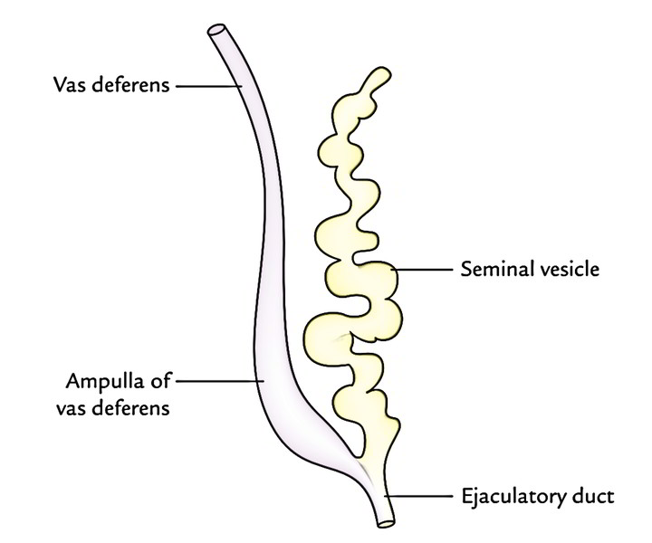
Ductus (Vas) Deferens
A muscular tube that transports sperm from the epididymis to the ejaculatory ducts. Its course includes:
- Originating from the deep inguinal ring.
- Passing superior to the ureters.
- Approaching the prostate medial to the seminal vesicles.

Female Internal Genital Organs
Uterus
The uterus is a hollow muscular organ essential for reproductive functions. It includes:
- Fundus: The top portion above the entry of the fallopian tubes.
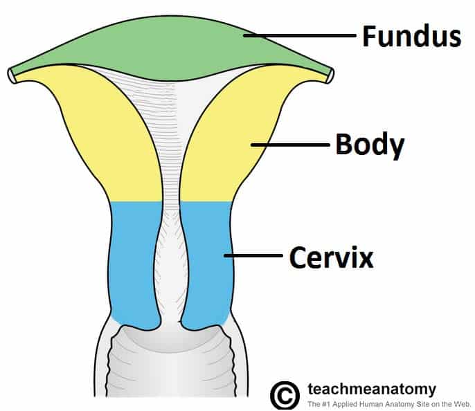
- Body: The central region.
- Cervix: The lower part that opens into the vagina.
Uterine (Fallopian) Tubes
These extend from the uterus to the ovaries, facilitating the passage of eggs. They include distinct sections:
- Isthmus: The narrow section connecting to the uterus.
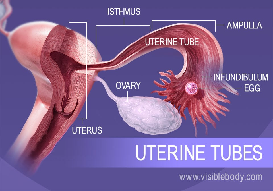
- Ampulla: The wider area where fertilization typically occurs.
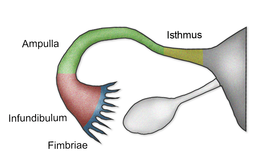
- Infundibulum: The funnel-shaped opening near the ovary, with finger-like projections called fimbriae.

Ovaries
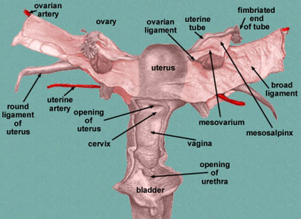
These are female gonads producing eggs and hormones. The ovaries are anchored by several ligaments:
- Suspensory Ligament: Contains ovarian vessels.
- Ovarian Ligament: Connects the ovary to the uterus.
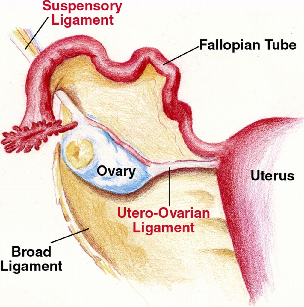
- Broad Ligament: A peritoneal fold that includes the mesosalpinx, mesovarium, and mesometrium.
Round Ligament of the Uterus
This ligament passes from the uterus to the labia majora via the inguinal canal, maintaining the anteverted position of the uterus.
Pelvic Vasculature
Arterial Supply
The major arterial supply to the pelvis includes:
- Common Iliac Arteries: Divide into external and internal iliac arteries.

- Internal Iliac Arteries: Branch into anterior and posterior divisions, supplying pelvic organs, perineum, and gluteal region.
/GettyImages-87377010-72ec77ac5a2745f086d00625d0d0d8f6.jpg)
- External Iliac Arteries: Continue as the femoral arteries to supply the lower limbs.
Venous Drainage
Pelvic venous drainage corresponds with arterial supply, including:
- Internal Iliac Veins: Drain into the common iliac veins.
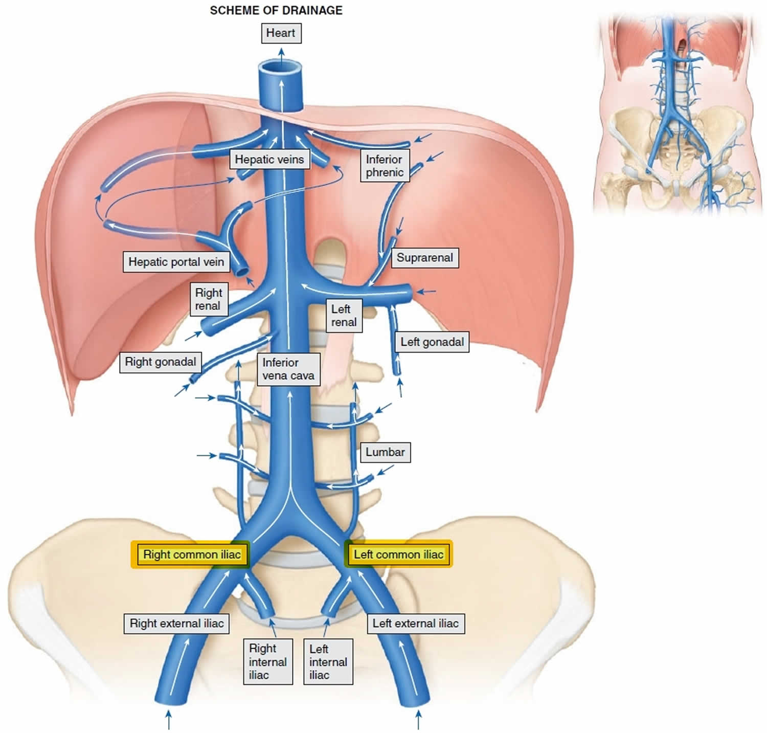
- External Iliac Veins: Continue as the femoral veins.
/GettyImages-87394488-bdb4ec80949c43519a5e2fed2e916ad6.jpg)
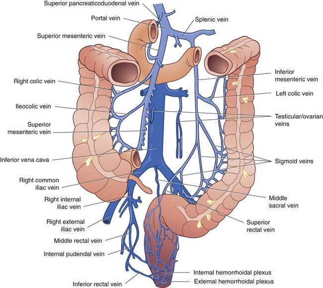
- Rectal Venous Plexuses: Include internal (superior rectal veins) and external (inferior rectal veins) leading to internal and external hemorrhoids respectively.
Lymphatic Drainage
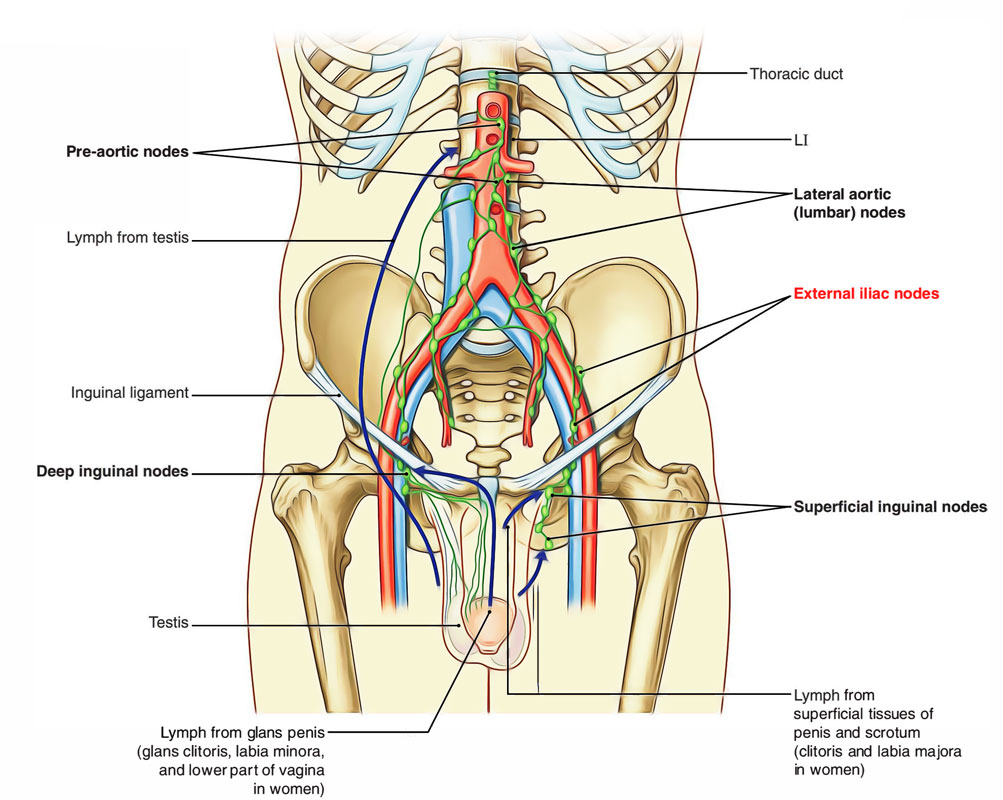
Pelvic lymph nodes include:
- Internal Iliac Nodes: Drain the pelvic organs.

- External Iliac Nodes: Drain the lower limb and anterior abdominal wall.
- Common Iliac Nodes: Drain into the lumbar (aortic) nodes.
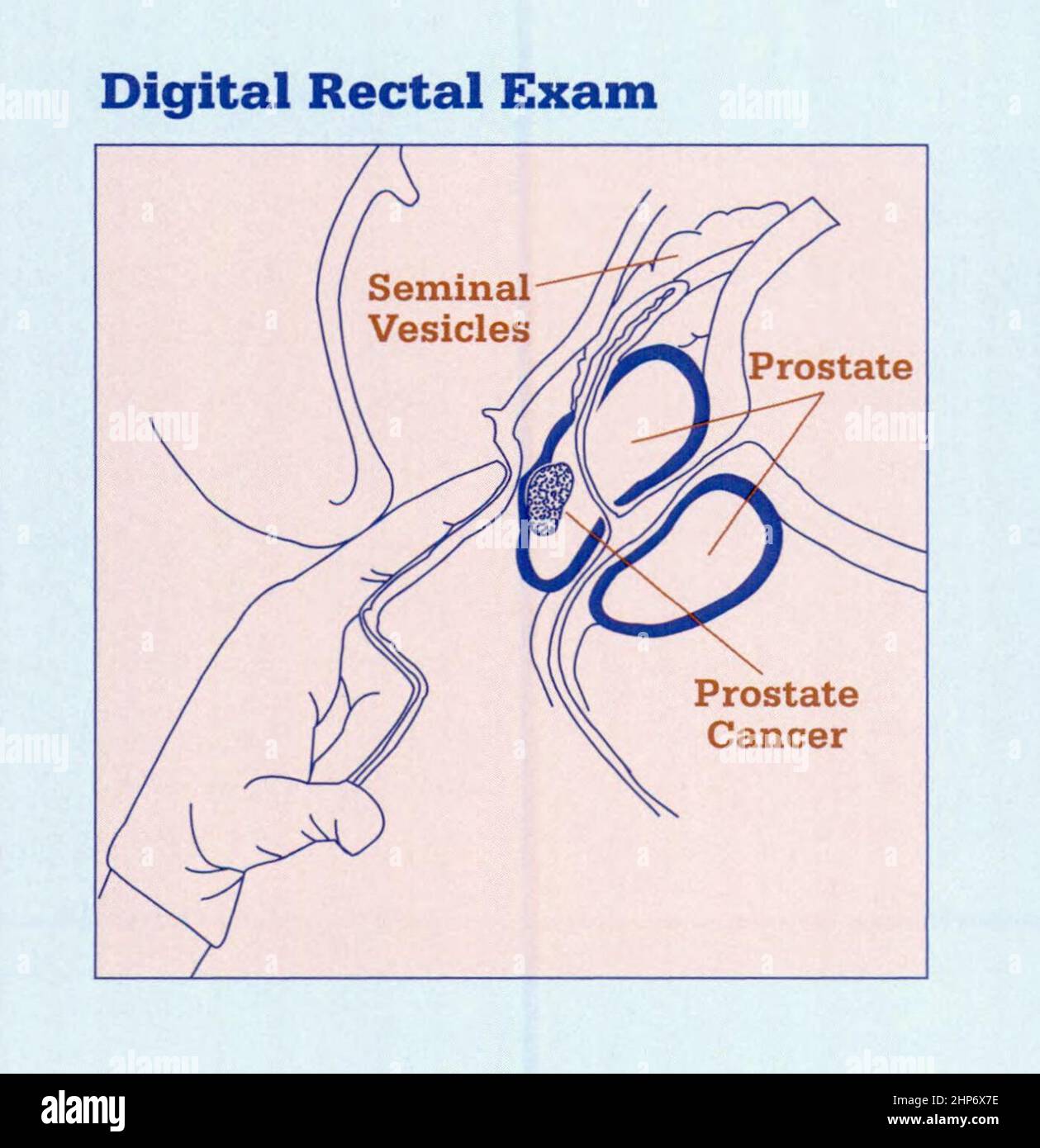
Rectal Digital Examination
A rectal examination in both males and females can evaluate:
- Prostate (in males): Check for enlargement or nodules.
- Cervix (in females): Evaluate its position and condition.
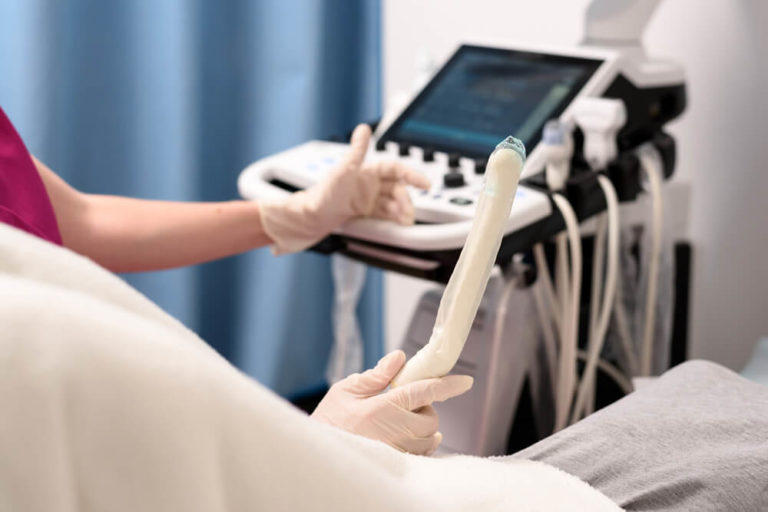
- Other Structures: Including the sacrum, coccyx, and ischial spines. Detection of enlarged lymph nodes or masses is also possible.
