Back and Spinal Cord
The Anatomy and Physiology of the Spinal Cord
Spinal Cord Overview
The spinal cord, a vital central nervous system (CNS) structure, extends from the foramen magnum at the skull base to the first or second lumbar vertebra (L1-L2) level in adults. It is a major conduit for transmitting neural signals between the brain and the rest of the body, facilitating motor control, sensory perception, and autonomic functions. The spinal cord is housed within the vertebral column and protected by cerebrospinal fluid (CSF), which cushions and nourishes it.
Anatomy of the Spinal Cord
1. Gross Anatomy

*Note from an M2: Identify the filum terminal, conus medullaris, and cauda equina. Know the order or the structures you will come in contact with if you do a lumbar puncture.
- Segments: It is divided into 31 segments, each giving rise to a pair of spinal nerves:
- Cervical: 8 segments (C1-C8)
- Thoracic: 12 segments (T1-T12)
- Lumbar: 5 segments (L1-L5)
- Sacral: 5 segments (S1-S5)
- Coccygeal: 1 segment (Co1)
- Conus Medullaris: The tapered end of the spinal cord, typically at the L1-L2 vertebral level.
- Cauda Equina: A bundle of lumbar and sacral nerve roots that extend beyond the conus medullaris within the vertebral canal.
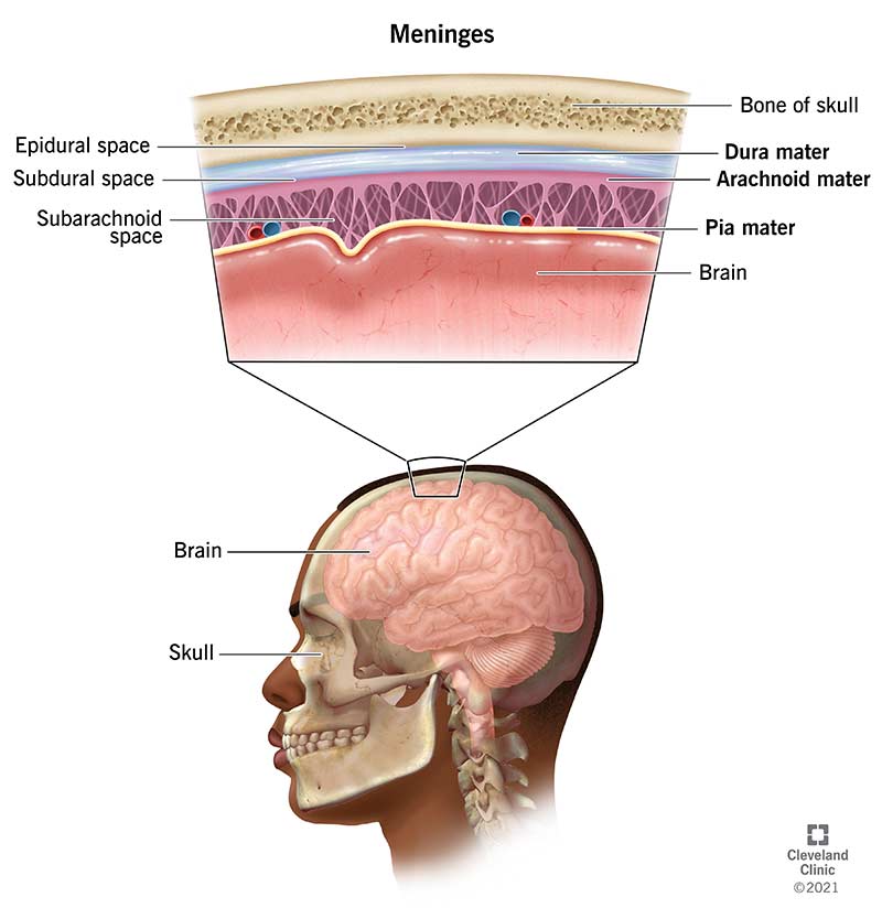
2. Meninges
- Dura Mater: The outermost, tough protective sheath.
- Arachnoid Mater: The middle, web-like layer filled with CSF.
- Pia Mater: The innermost layer is closely adhered to the surface of the spinal cord.
Internal Structure
1. Gray Matter

- Shape and Location: The gray matter is centrally located, forming an H-shaped or butterfly structure in cross-section. It contains neuronal cell bodies, dendrites, and unmyelinated axons.
- Regions:
- Dorsal (Posterior) Horn: Contains sensory neurons that receive afferent signals from the body.
- Ventral (Anterior) Horn: Contains motor neurons that send efferent signals to muscles.
- Lateral Horn: Present in the thoracic and upper lumbar segments; contains neurons of the autonomic nervous system.
2. White Matter

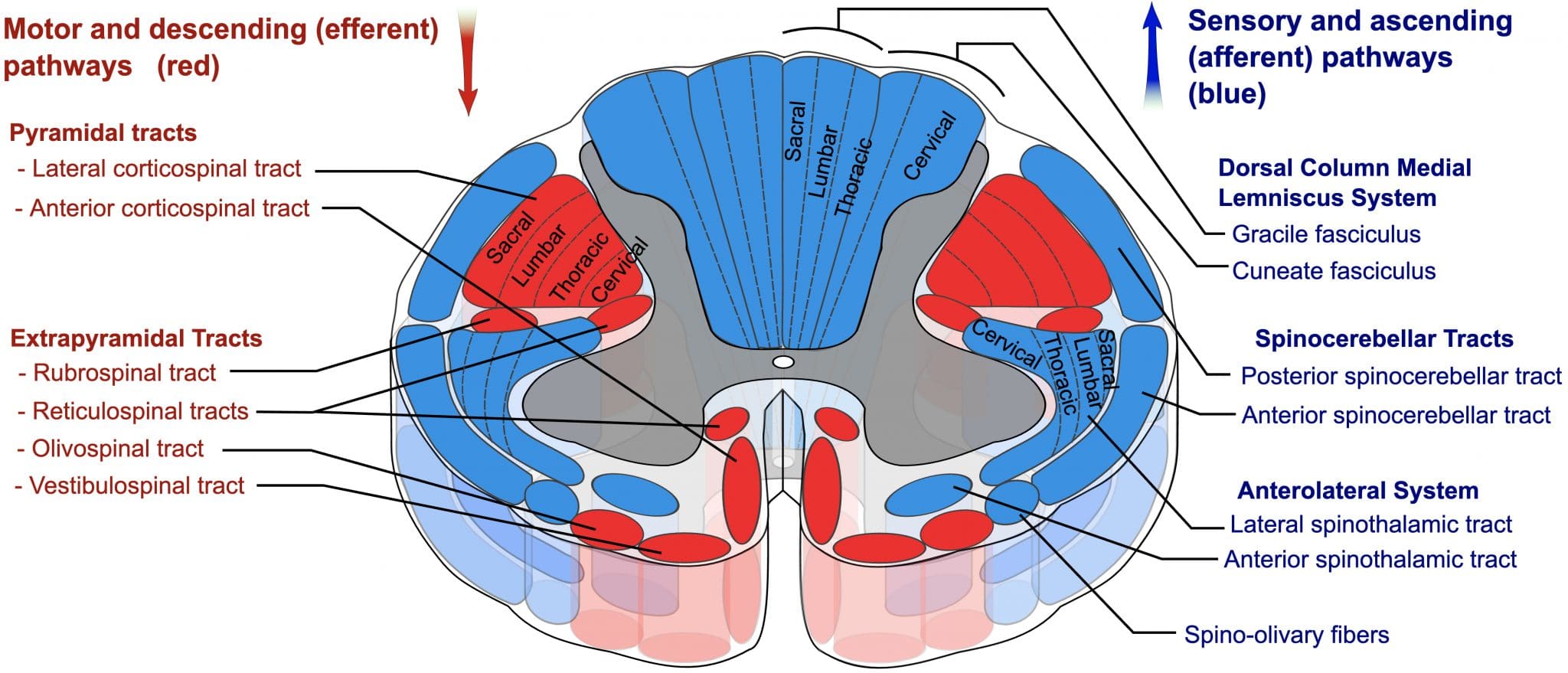
- Composition: Surrounding the gray matter, the white matter consists of myelinated axons grouped into tracts.
Here's a silly AI generated song to help you remember the content:
For Human Structures, you don't need to know the tracts, but they will come up endlessly in Brain and Behavior, so everything below this point is just a preview of what you'll learn later on.
- Tracts:
- Ascending Tracts: Carry sensory information from the periphery to the brain. Key tracts include:
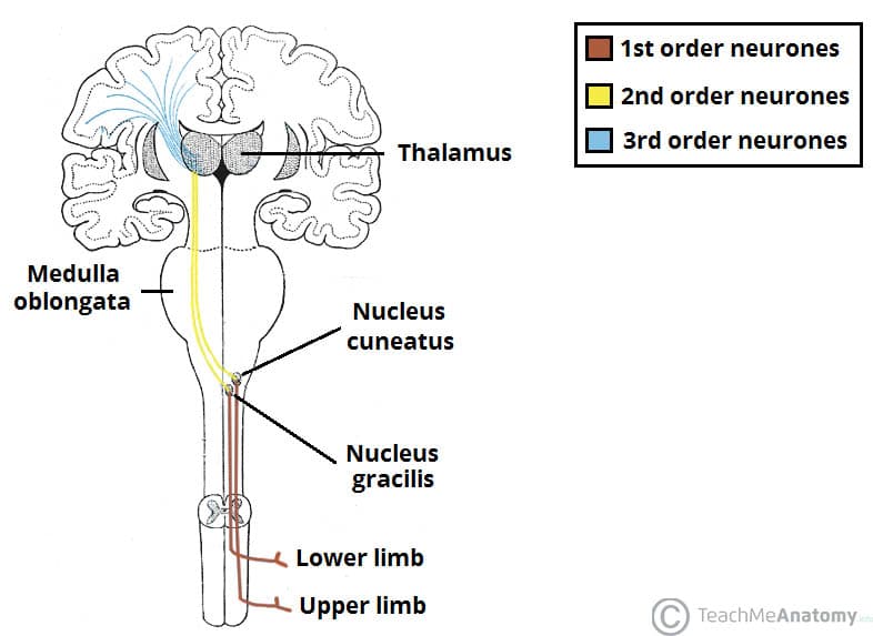
- Dorsal Columns: Fine touch, vibration, and proprioception.
- Spinothalamic Tract: Pain, temperature, and crude touch.
- Spinocerebellar Tract: Proprioceptive information to the cerebellum.
- Descending Tracts: Convey motor commands from the brain to the periphery. Key tracts include:
- Corticospinal Tract: Voluntary motor control.
- Reticulospinal and Vestibulospinal Tracts: Posture and balance.
- Rubrospinal Tract: Motor control, particularly of the upper limbs.
*Again, this is just a sneak peek at brain and behavior, so you're exposed to it. Don't worry about this too much now. Just know it exists.
- Ascending Tracts: Carry sensory information from the periphery to the brain. Key tracts include:

Functional Anatomy
1. Sensory Pathways
- Dorsal Column-Medial Lemniscal Pathway: Responsible for transmitting delicate touch, vibration, and proprioception.
- Anterolateral System: Comprising the spinothalamic tract, this system transmits pain, temperature, and crude touch.
2. Motor Pathways
- Pyramidal Tracts (Corticospinal Tracts): Major pathways for voluntary motor control. They originate in the cerebral cortex and synapse with lower motor neurons in the spinal cord.
- Extrapyramidal Tracts: Include the reticulospinal, vestibulospinal, rubrospinal, and tectospinal tracts, which are involved in involuntary and automatic control of muscles .

Clinical Correlation: Spinal Cord Injuries
Common Syndromes:
- Central Cord Syndrome: Typically results from cervical spinal cord damage, leading to greater weakness in the upper limbs than in the lower limbs.
- Anterior Cord Syndrome: Damage to the anterior spinal artery, affecting the anterior two-thirds of the spinal cord, leading to motor loss and loss of pain and temperature sensation.
- Brown-Séquard Syndrome: Hemisection of the spinal cord, leading to ipsilateral motor loss and contralateral sensory loss.

Spinal Cord Vascular Supply
The spinal cord receives blood supply from:
- Anterior Spinal Artery: Supplies the anterior two-thirds of the spinal cord.
- Posterior Spinal Arteries: Supply the posterior one-third of the spinal cord.
- Radicular Arteries: Branch from segmental arteries and reinforce the spinal arteries.

