Breasts and Pectoral Muscles
Skeleton of the Pectoral Region
Notes from an M2:
It's important to know what is palpable, which numbers of ribs are true/false etc, and the different areas of the chest cavity.
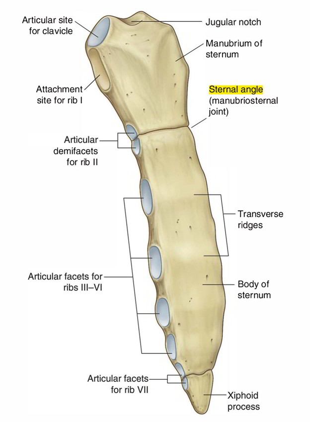
Manubrium
- Description: The upper part of the sternum.
- Function: Provides attachment for the clavicles and the first pair of ribs.
Sternal Angle of Louis
- Description: A palpable landmark where the manubrium joins the body of the sternum.
- Location: Level of the second rib's attachment.
- Significance: A critical anatomical reference point.

Sternum
- Description: A long, flat bone forming the middle portion of the anterior wall of the thorax.
- Function: Provides attachment points for the ribs via costal cartilage.

Ribs
- True Ribs (1-7): Direct anterior attachment to the sternum via costal cartilage.
- False Ribs (8-10): Attached indirectly to the sternum via the cartilage of the rib above.
- Floating Ribs (11-12): No anterior attachment to the sternum.
Clavicle and Scapula
- Components: Clavicle (collarbone) and scapula (shoulder blade).
- Function: Make up the shoulder girdle, part of the appendicular skeleton.
Veins of the Anterior Wall - Superficial and Deep
Key Veins:
- Axillary Vein: Continuation of the basilic vein; drains blood from the upper limb.
- Cephalic Vein: Superficial vein running along the lateral side of the arm.
- Internal Thoracic Vein: Drains into the brachiocephalic veins.
- Lateral Thoracic Vein: Drains the lateral aspects of the thorax.
- Anterior Intercostal Veins: Drain blood from the intercostal spaces.

Mammary Gland
Components
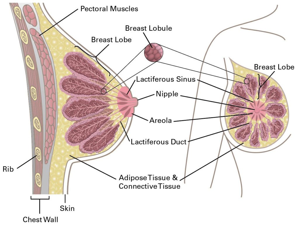
- Suspensory Ligaments (Cooper's Ligaments): Support the breasts by connecting the skin to the underlying pectoral fascia.
- Areolar Glands: Sebaceous glands in the areola surrounding the nipple.
- Axillary Tail: Extension of the breast tissue into the armpit (axilla).
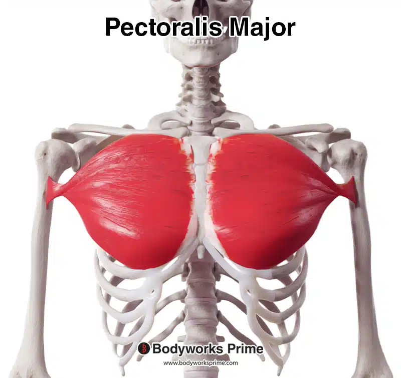
- Pectoralis Major Muscle: Lies deep to the pectoral fascia and supports the breast.
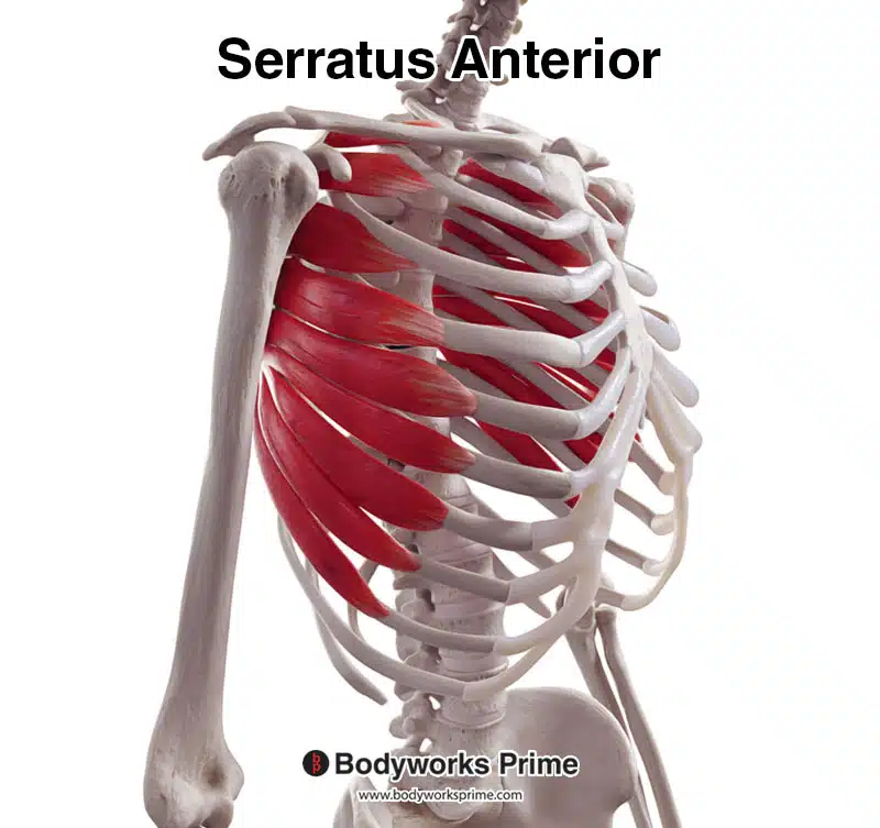
- Serratus Anterior Muscle: Located on the side of the thorax, aiding in respiration and shoulder movement.

Structure
- Lobes and Lobules: The mammary gland contains 15-20 lobes, each consisting of several lobules.
- Lactiferous Ducts: Transport milk from the lobules to the nipple.
- Nipple and Areola: The nipple is surrounded by the pigmented areola.
- Lactiferous Sinus: Expanded portion of the duct just beneath the nipple.
- Fat: Provides shape and volume to the breast.
Hormonal Control
- Estrogen: Promotes growth of ducts and periductal connective tissue.
- Progesterone: Stimulates growth and differentiation of lobules.
- Prolactin: Essential for gland development and milk production.
- Oxytocin: Facilitates milk ejection during breastfeeding.

Clinical Significance
- Gynecomastia: Development of breast tissue in males due to hormonal imbalances.
- Fibrocystic Changes: Formation of fluid-filled cysts and fibrous tissue, causing the breast to feel lumpy or tender.
- Carcinomas: Tumors originating from epithelial tissue; most common type of breast cancer.
- Sarcomas: Tumors from mesodermal tissues.
- Lymphatic Drainage: Critical in the spread of cancerous cells; lack of encapsulation makes mammary glands susceptible to metastasis.

Arteries Supplying the Mammary Glands
- Internal Thoracic Artery: Supplies the medial part of the breast.
- Lateral Thoracic Artery: Supplies the lateral part of the breast.
- Posterior Intercostal Arteries: Contribute to the blood supply of the breast.
- Thoracoacromial Artery: Involved in the vascularization of the breast tissue.

Lymphatic Drainage
*NOTE: Just knowing the general lymphatic drainage pattern suffices for m1.
- Lymphatic System: Drains excess fluid and plasma proteins from breast tissue and transports them to major veins.
- Lymph Nodes: Filter fluid; vital for cancer diagnosis and treatment.
- Axillary Lymph Nodes Include the apical (subclavian) and various other groups, such as the central, pectoral, subscapular, and lateral nodes. Cancer can spread through these nodes, highlighting the importance of lymphatic drainage in breast pathology.
:watermark(/images/watermark_5000_10percent.png,0,0,0):watermark(/images/logo_url.png,-10,-10,0):format(jpeg)/images/overview_image/636/j7MyyazXblAZKZJUlQMWyw_18_the-lymphatics-of-the-female-breast_english.jpg)
Muscles of the Pectoral Region
Pectoralis Major Muscle
- Origin: Clavicular head from the anterior medial clavicle, sternal head from the anterior sternum, and the superior six costal cartilages.
- Insertion: Crest of the greater tubercle of the humerus.
- Innervation: Lateral and medial pectoral nerves.
- Function: Adducts and medially rotates the humerus; draws the scapula anteriorly and inferiorly.

Pectoralis Minor Muscle
- Origin: Third to fifth ribs.
- Insertion: Coracoid process of the scapula.
- Innervation: Medial pectoral nerve.
- Function: Stabilizes the scapula by drawing it inferiorly and anteriorly against the thoracic wall.
:background_color(FFFFFF):format(jpeg)/images/article/pectoralis-minor-muscle/RByPN6lyb7aFQEbwF4Z2qQ_pectoralis_minor_muscle.png)
Subclavius Muscle
- Origin: First rib and its cartilage.
- Insertion: Inferior surface of the middle third of the clavicle.
- Innervation: Nerve to subclavius (C5, C6).
- Function: Anchors and depresses the clavicle.

Serratus Anterior Muscle
- Origin: Ribs 1-8.
- Insertion: Anterior surface of the medial border of the scapula.
- Innervation: Long thoracic nerve.
- Function: Protracts, secures, and rotates the scapula.

Vasculature of the Pectoral Region
Arteries of the Upper Limb
- Axillary Artery and Its Branches:
- First Part: Superior thoracic artery.
- Second Part: Thoracoacromial artery and lateral thoracic artery.
- Third Part: Subscapular artery, anterior and posterior circumflex humeral arteries.

