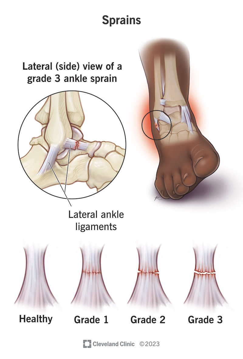
Tibiofibular Joints and Skeleton of the Foot
Tibiofibular Joints and Skeleton of the Foot
Tibiofibular Joints
The tibiofibular joints are composed of three parts, each with its distinct characteristics and functional significance:
- Superior Tibiofibular Joint: This is a plane synovial joint located between the lateral condyle of the tibia and the head of the fibula.
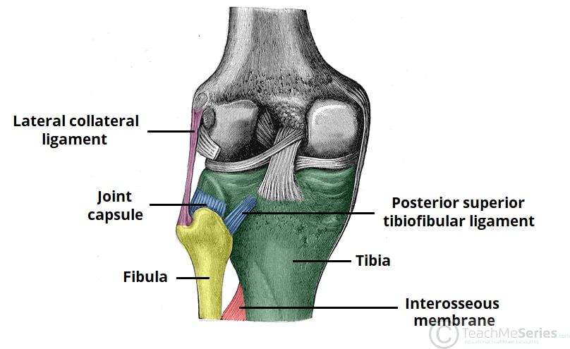
- Middle Tibiofibular Joint: Known as syndesmosis, this is a fibrous joint where the interosseus membrane connects the shafts of the tibia and fibula along their length.
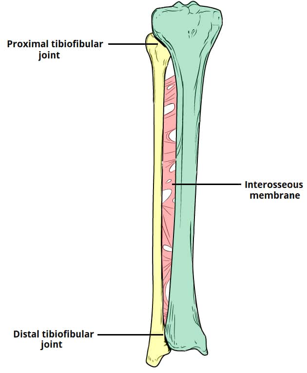
- Inferior Tibiofibular Joint: Another syndesmosis, this joint connects the distal ends of the tibia and fibula, contributing to the stability of the ankle.
:background_color(FFFFFF):format(jpeg)/images/library/13295/Tibiofibular_joints.png)
Anatomy of the Tibia and Fibula
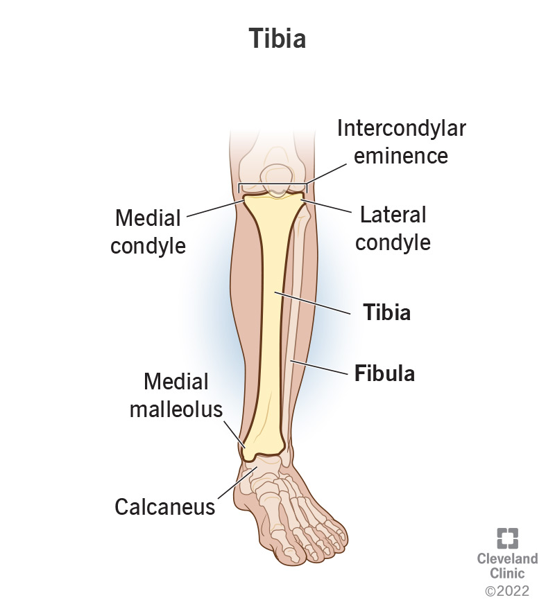
Tibia: Carries body weight to the foot.
- Anterior Border of Tibia
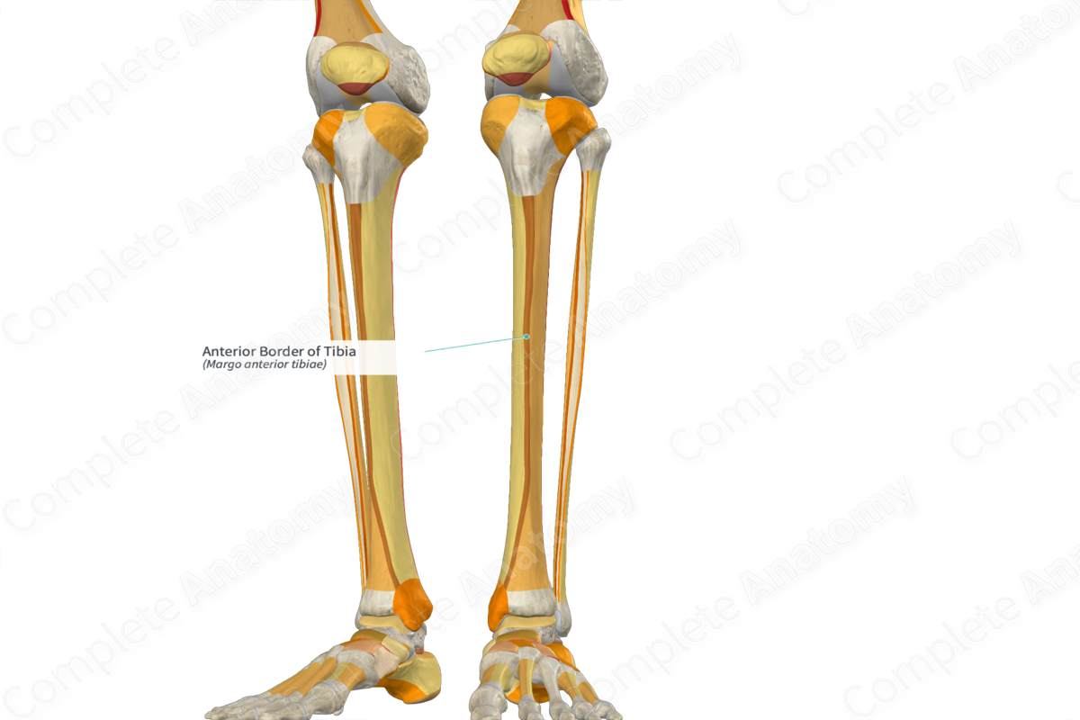
- Interosseus Border of Tibia
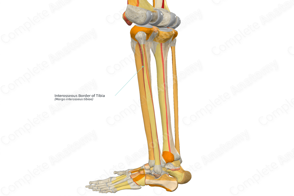
- Anterior Border of Tibia
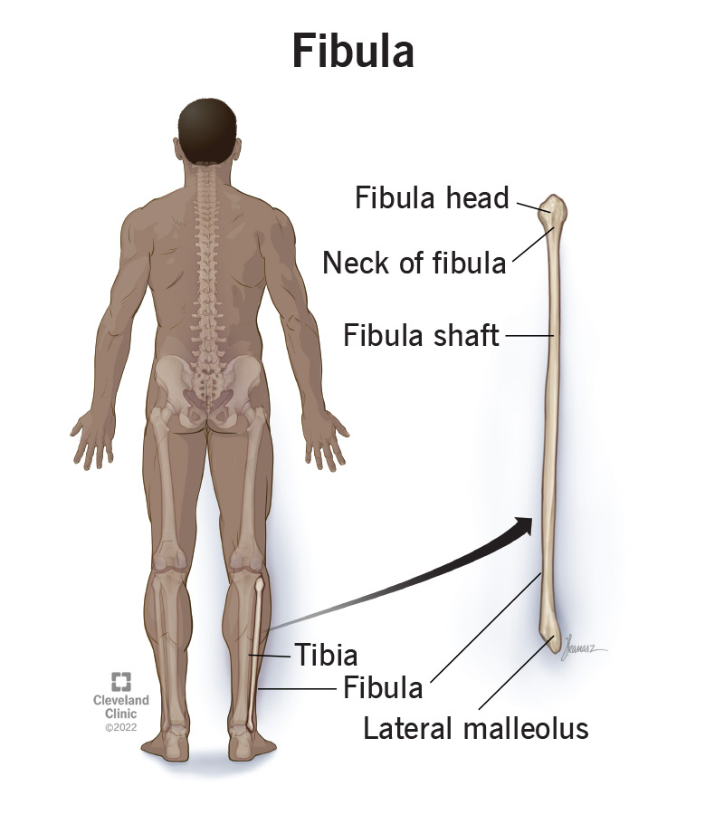
Fibula: Provides muscle attachment.
- Interosseus Border of Fibula

- Interosseus Border of Fibula
Key ligaments associated with these bones:
- Anterior Tibiofibular Ligament
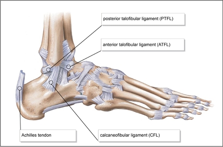
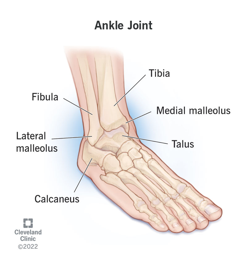
- Lateral Malleolus
- Medial Malleolus
Skeleton of the Foot
The foot is composed of several bones that can be categorized into three groups: tarsal bones, metatarsals, and phalanges. Each of these bones plays an essential role in the structural integrity and movement of the foot.
Tarsal Bones
- Calcaneus: The largest tarsal bone that transmits most of the weight from the talus to the ground.
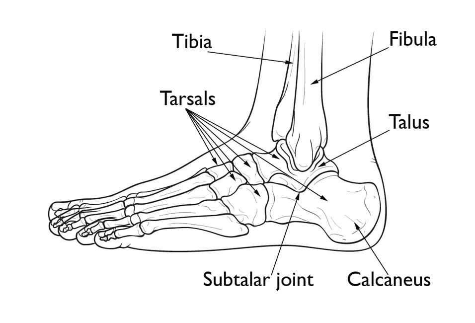
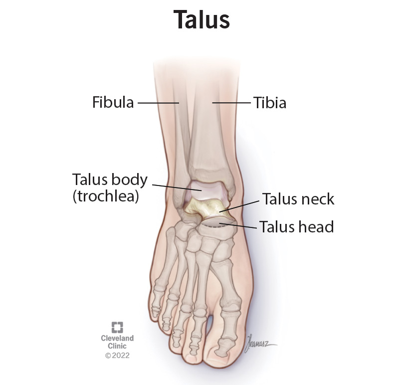
- Talus: The only tarsal bone that articulates with the leg bones; no muscle attaches to the talus.
- Parts: Trochlea, Neck, Head
- Parts: Trochlea, Neck, Head
- Navicular
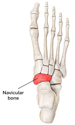
- Cuboid
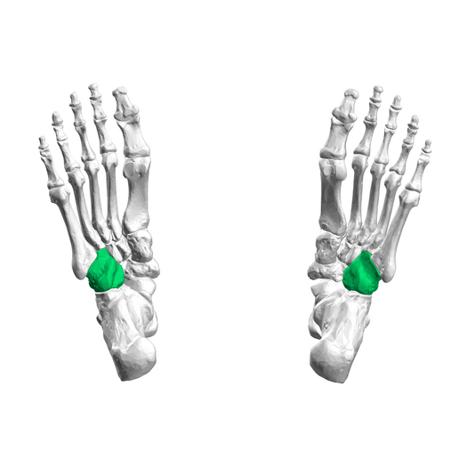
- Medial Cuneiform
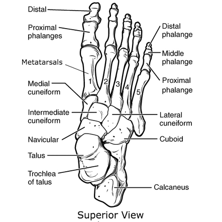
- Intermediate Cuneiform

- Lateral Cuneiform

Metatarsals (5)
- The long bones in the foot, numbered I to V, from the medial (big toe) to the lateral (little toe) sides.

- Base, Shaft, Head
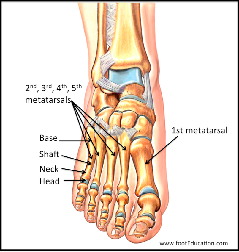
- Base, Shaft, Head
Phalanges
- Proximal Phalanges (5)
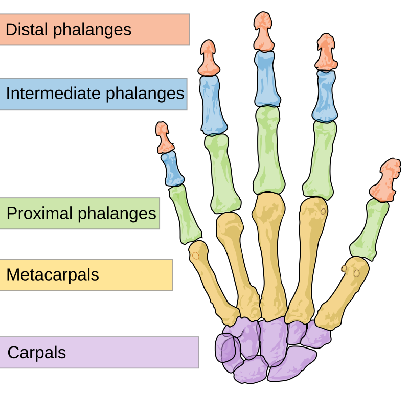
- Middle Phalanges (4)
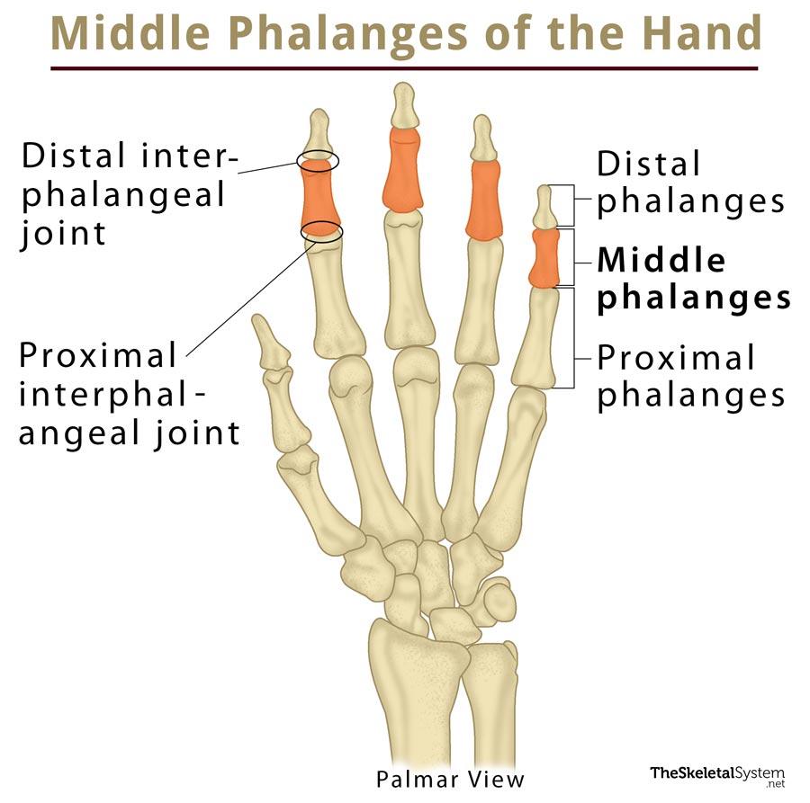
- Distal Phalanges (5)
Arches of the Foot
The foot has two main arches, which act as shock absorbers:
- Medial Longitudinal Arch: Formed by the calcaneus, talus, navicular, medial cuneiform, and the 1st metatarsal.
- Lateral Longitudinal Arch: Made up of the calcaneus, cuboid, and the 5th metatarsal.

- Transverse Arch: Composed of the cuboid and the three cuneiforms.

Ligaments and Tendons
Key ligaments and tendons supporting the foot:
- Plantar Calcaneonavicular (Spring) Ligament
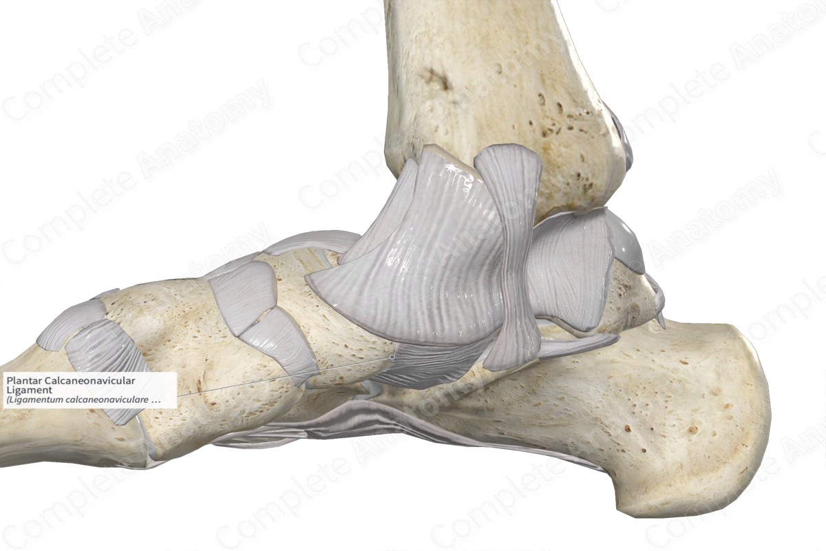
- Long Plantar Ligament
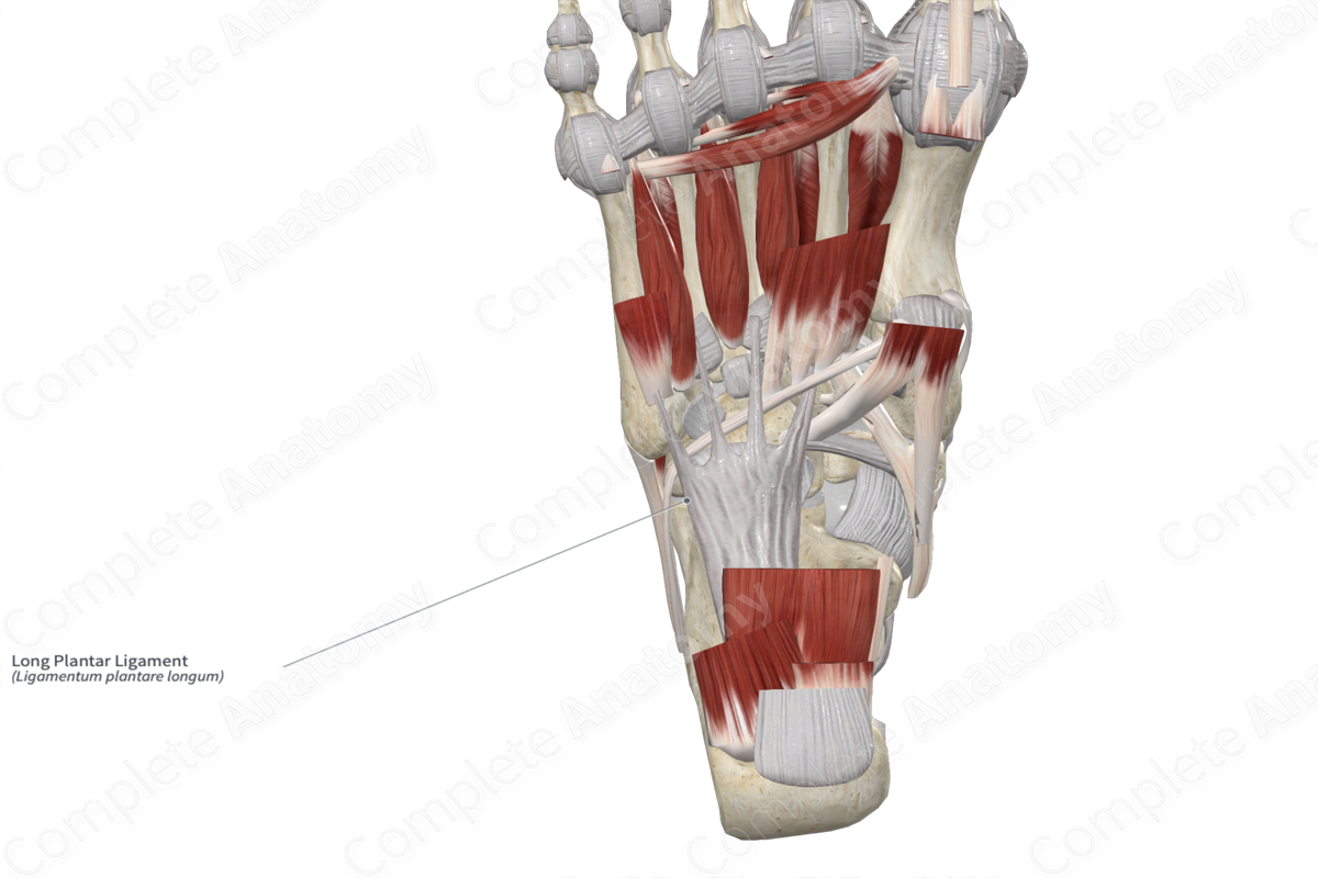
- Short Plantar Ligament

- Tibialis Anterior Tendon

- Tibialis Posterior Tendon
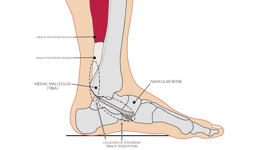
- Fibularis Longus Tendon

- Fibularis Brevis Tendon
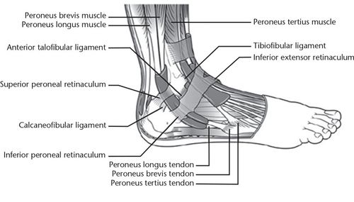
Ankle Joint
Lateral View
The ankle joint consists of several ligaments that provide stability:
- Posterior Talofibular Ligament

- Calcaneofibular Ligament
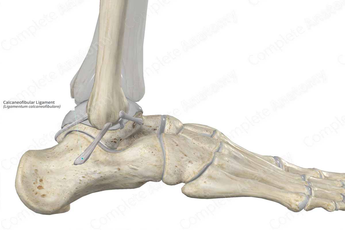
- Anterior Talofibular Ligament

Medial View
The medial ligaments are stronger and help in stabilizing the joint during eversion, preventing dislocation:
- Deltoid Ligament
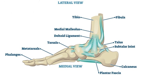
- Post. Tibiotalar
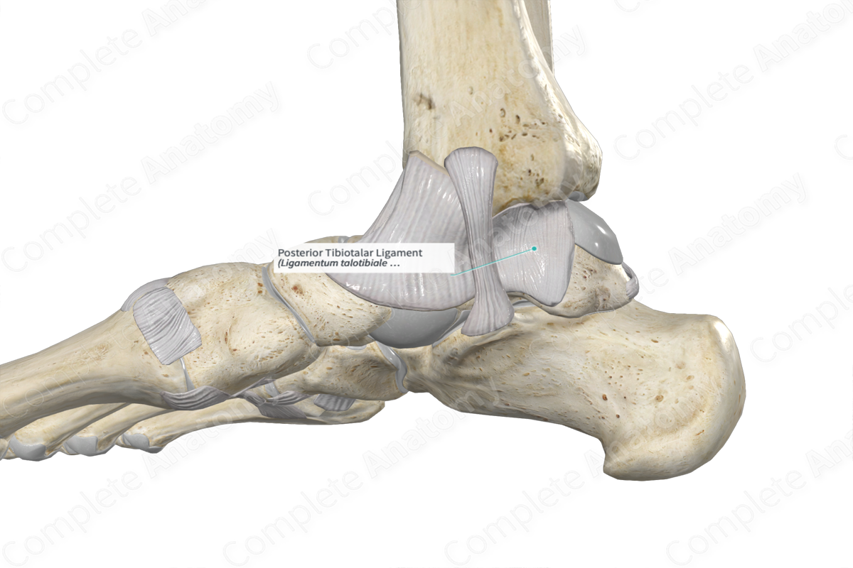
- Tibiocalcaneal
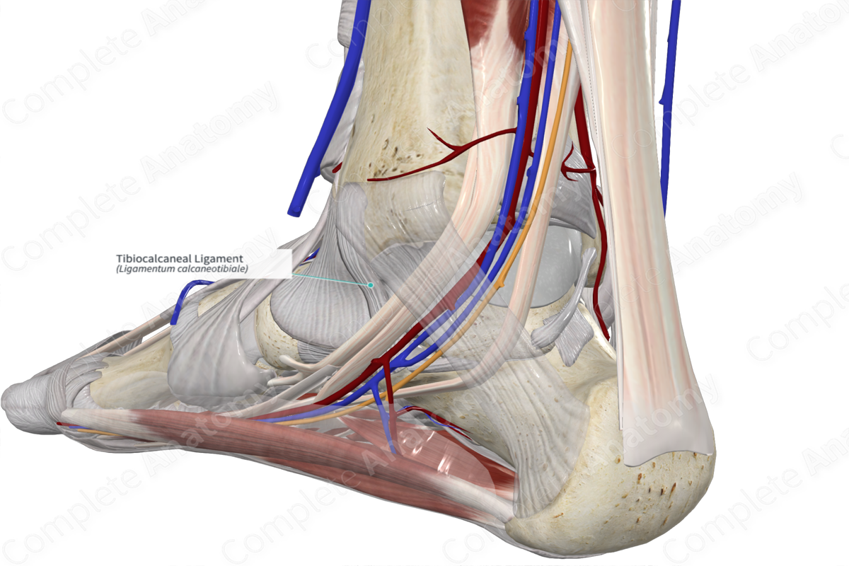
- Tibionavicular
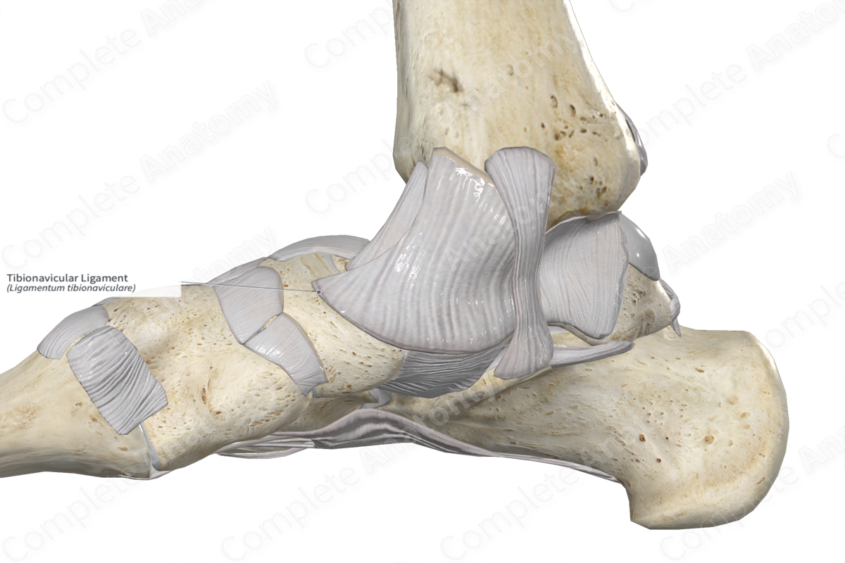
- Ant. Tibiotalar
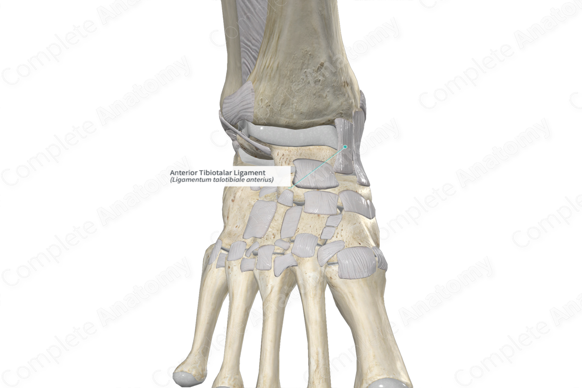
- Post. Tibiotalar
Movements of the Ankle Joint
The primary movements at the ankle joint are dorsiflexion and plantar flexion:
- Dorsiflexion: Performed by the muscles of the anterior compartment.

- Plantar Flexion: Performed by the muscles of the posterior compartment.

Injuries
- Ankle Sprains: Often involve lateral ligament sprains with foot inversion.
- Pott Fracture: Dislocation of the ankle during forcible eversion.
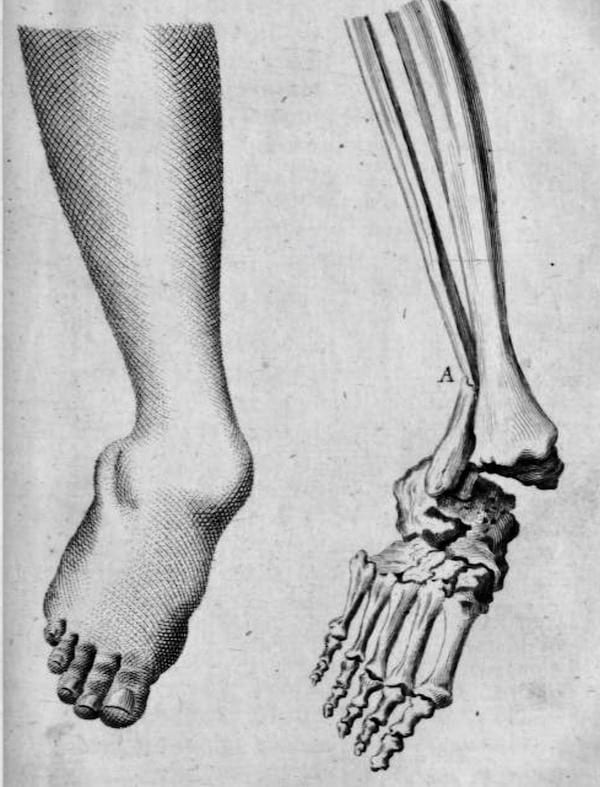
Joints Involved in Foot Movements
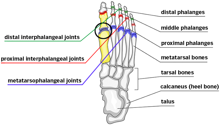
- Interphalangeal Joints
:background_color(FFFFFF):format(jpeg)/images/library/13136/Wviyodz2L9s0cCTIK65hAA_Ligamentum_coracoclaviculare_01.png)
- Metatarsophalangeal Joints
- Talocalcaneonavicular Joint
:background_color(FFFFFF):format(jpeg)/images/library/13267/bOrEuyFKqxOJq6WjmWew5A_talonavicular_part_of_talocalcaneonavicular_joint.png)
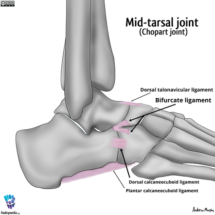
- Transverse Tarsal Joint
:background_color(FFFFFF):format(jpeg)/images/library/13309/Transverse_tarsal_joint.png)
- Calcaneocuboid Joint
- Tarsometatarsal Joints

- Subtalar Joint

Muscles of the Foot and Ankle
Anterior Compartment
- Tibialis Anterior
:background_color(FFFFFF):format(jpeg)/images/library/12972/ssEatJ6boVqj60odURIiQ_S6zoMopRSV_M._tibialis_anterior_NN_2.png)
- Extensor Digitorum Longus
:background_color(FFFFFF):format(jpeg)/images/library/12923/Extensor_digitorum_longus_muscle.png)
- Extensor Hallucis Longus
:background_color(FFFFFF):format(jpeg)/images/library/12862/xcwbVfplx1uRe1WwVe8Bw_uu5Wqf8cJG_M._extensor_hallucis_longus_NN_2.png)
- Fibularis Tertius (sometimes absent)
:background_color(FFFFFF):format(jpeg)/images/library/13168/Sn6nEzGnLI9n4zVfKtrjw_Fibularis_tertius.png)
Lateral Compartment
- Fibularis Longus
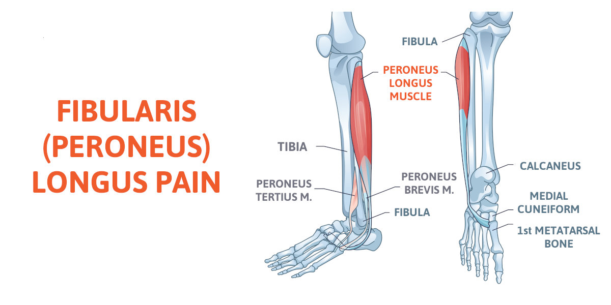
- Fibularis Brevis
:background_color(FFFFFF):format(jpeg)/images/library/12907/NF2DldEgb3o6tsP1c70u5Q_YWK3dQxWJL_M._peroneus_brevis_NN_1.png)
Posterior Compartment
Superficial Muscles
- Plantaris

- Gastrocnemius
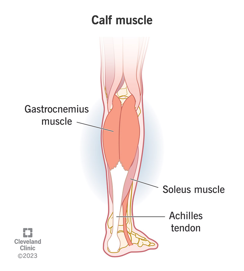
- Soleus

Deep Muscles
- Popliteus

- Flexor Digitorum Longus
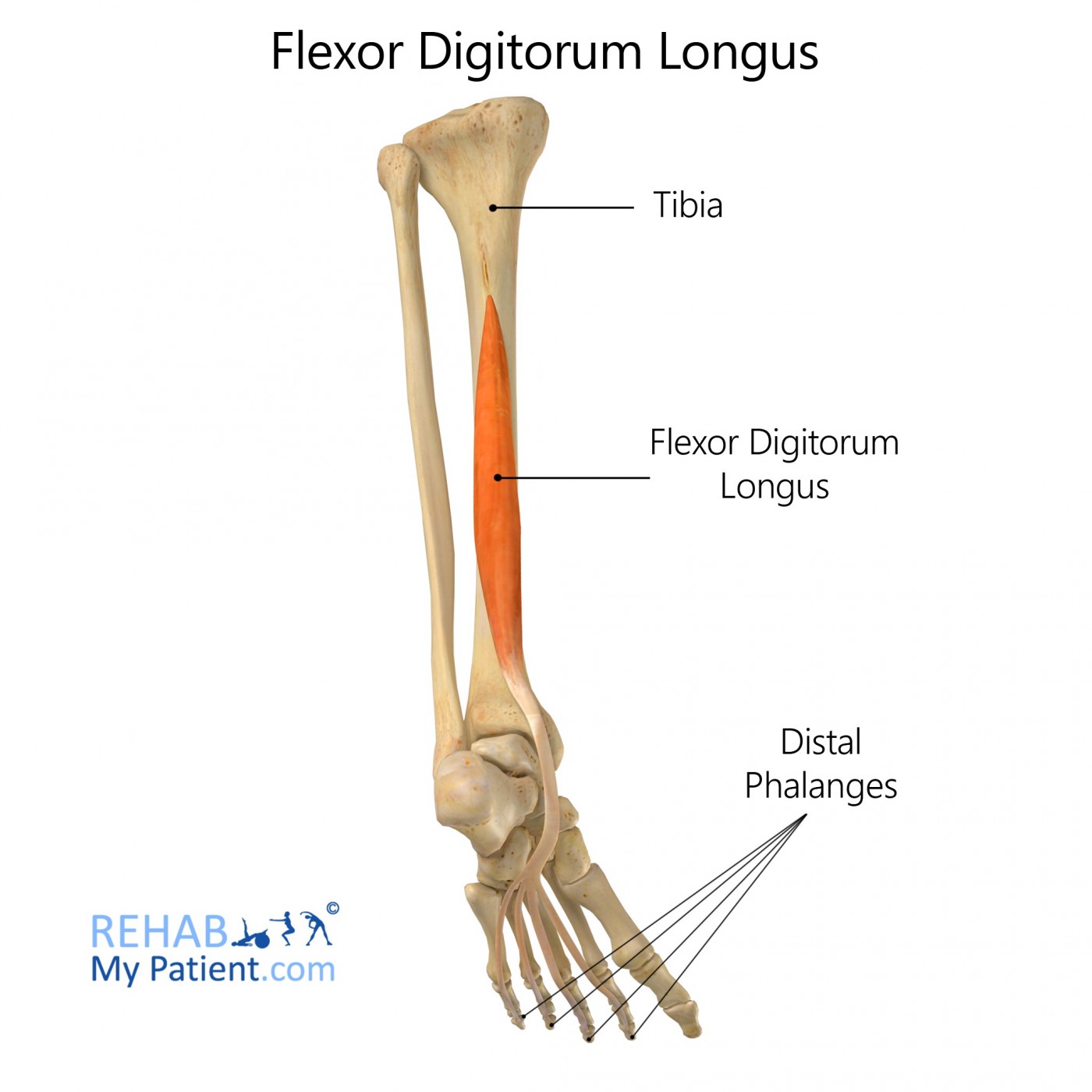
- Tibialis Posterior

- Flexor Hallucis Longus

Dorsal Muscles
- Extensor Digitorum Brevis
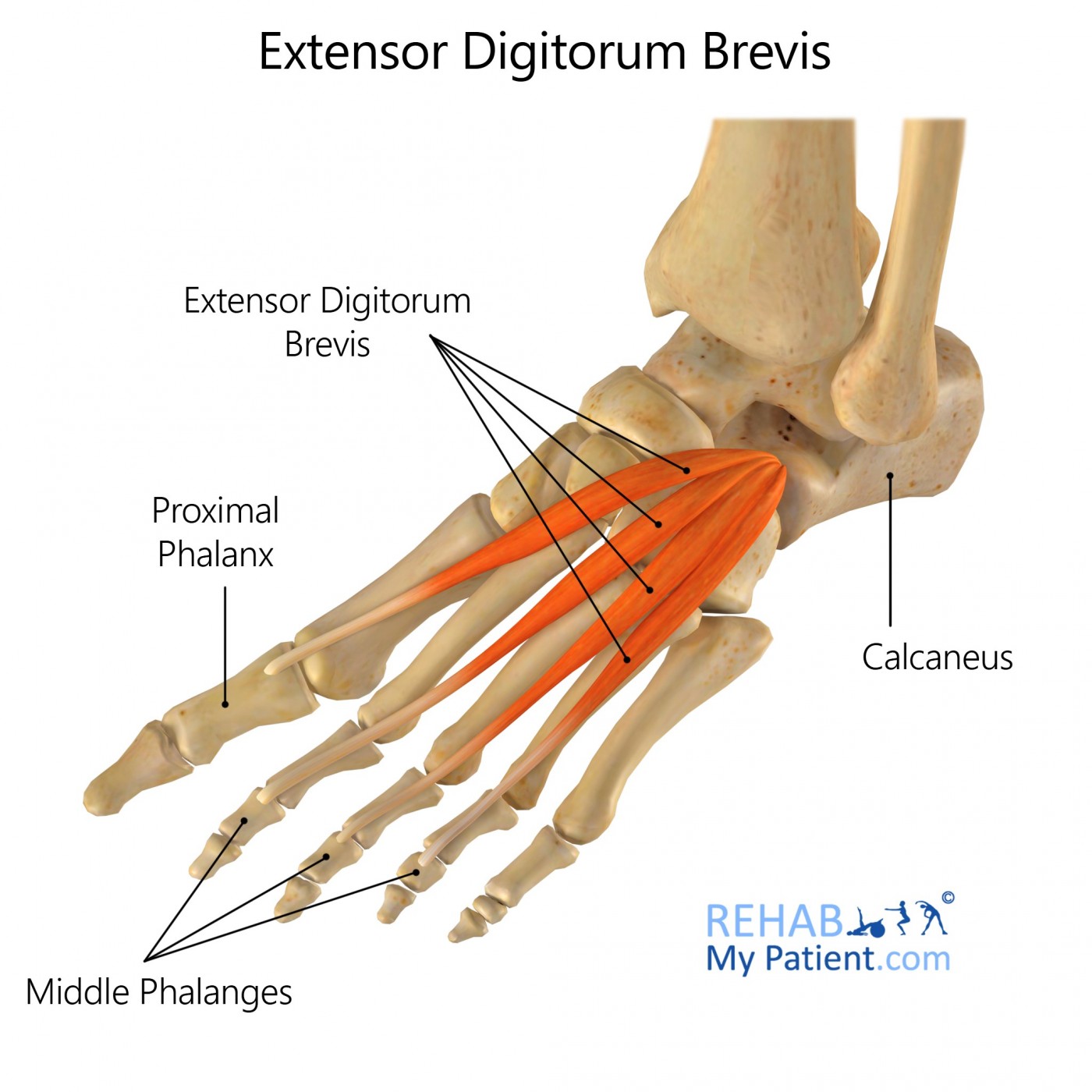
- Extensor Hallucis Brevis
:background_color(FFFFFF):format(jpeg)/images/library/12928/sdOjrUknu31WtEughpzQzA_cnWJNXDgxJ_M._extensor_hallucis_brevis_2.png)
Nerves and Arteries
Anterior Compartment
- Deep Fibular Nerve: Supplies the muscles of the anterior leg and dorsum of the foot.
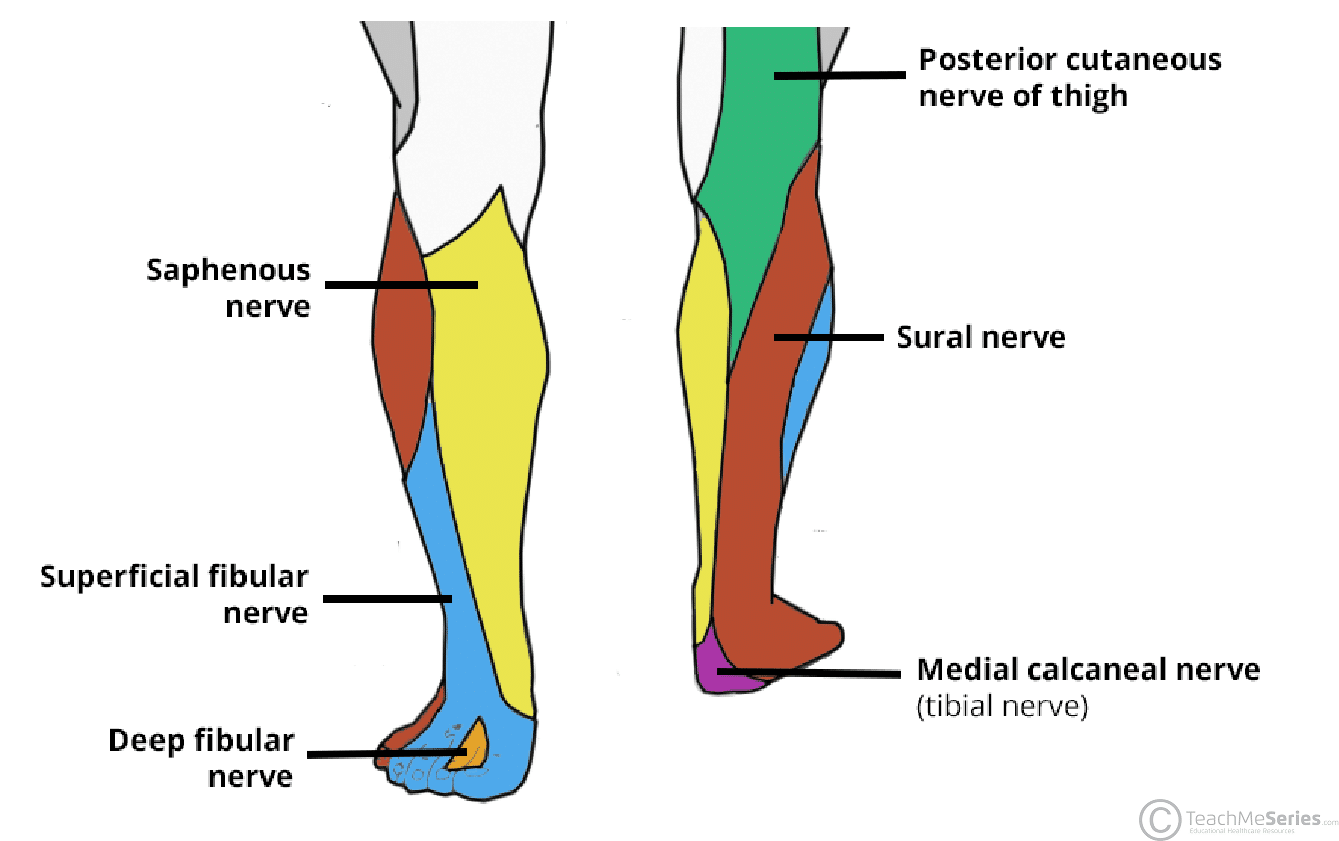
- Anterior Tibial Artery: Passes through a gap in the interosseous membrane.
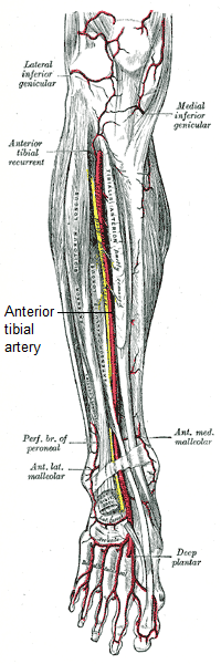
- Dorsalis Pedis Artery

Lateral Compartment
- Superficial Fibular Nerve: Supplies the muscles of the lateral compartment.

Posterior Compartment
- Tibial Nerve
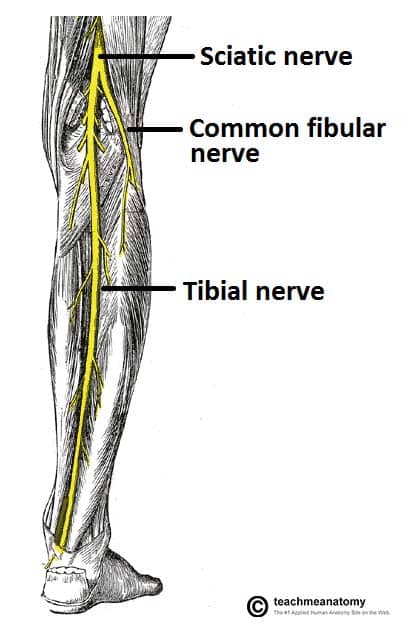
- Posterior Tibial Artery: With branches forming the fibular and perforating arteries.

- Fibular Artery

