Anatomy of the Neck and Related Structures
Anatomy of the Neck and Related Structures
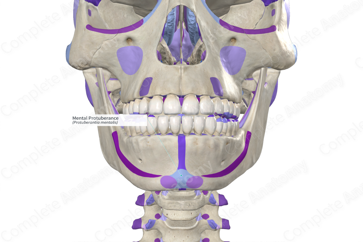
Mental Protuberance
The mental protuberance is the prominence at the front of the mandible (lower jawbone) forming the chin.
Mandible
The mandible, also known as the lower jaw or jawbone, is the largest, strongest, and lowest bone in the human face. It holds the lower teeth in place.

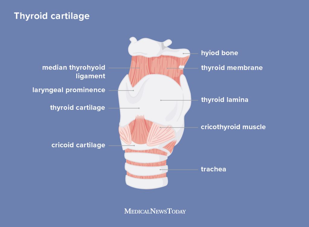
Thyroid Cartilage
The thyroid cartilage is the largest cartilage of the larynx and sits in front of the larynx and above the thyroid gland. It forms the Adam's apple.
Root of Neck
The root of the neck is the area at the junction of the thorax and the neck. It includes many vital structures like the great vessels (arteries and veins), parts of the brachial plexus, and the apexes of the lungs.

Muscular Structures (Mm)
- Digastric Muscle (Digastos): This muscle comprises two muscular bellies joined by an intermediate tendon, aiding in the movement of the jaw.
- Sternocleidomastoid Muscle (SCM): Extends from the sternum and clavicle to the mastoid process of the temporal bone, playing a key role in head movement and flexion of the neck.
:background_color(FFFFFF):format(jpeg)/images/article/digastric-muscle/34cPM1trZDDMFCSfr4bKdA_dkNB2edR8ILWCaohI4v5Sw_digastric.png)
:background_color(FFFFFF):format(jpeg)/images/article/sternocleidomastoid-muscle/N21GRSiWKOf8BQlrcQhA_hbimApe3IQfTimfNbNgnBw_sternocleido_mastoid.png)
Nerves
- Hypoglossal Nerve (CN XII): Innervates the muscles of the tongue.
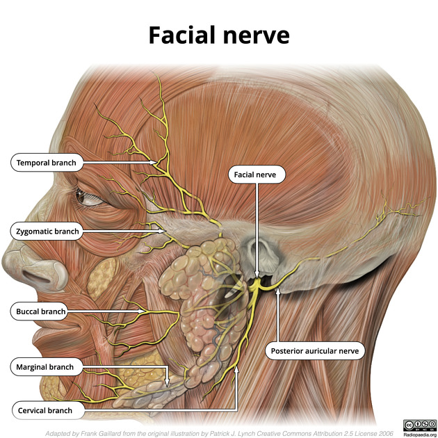
- Facial Nerve (CN VII): Controls the muscles of facial expression.
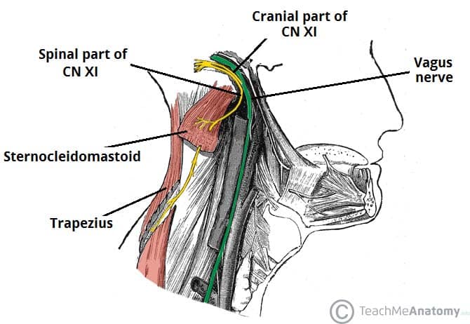
- Spinal Accessory Nerve (CN XI): Supplies the sternocleidomastoid and trapezius muscles.
- Ansa Cervicalis: A loop of nerves that supplies the infrahyoid (strap) muscles.
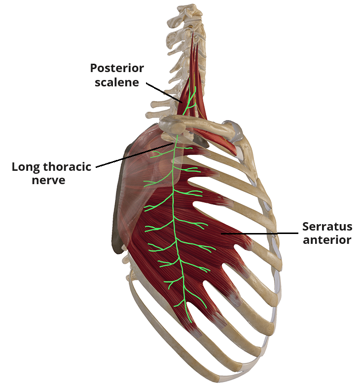
- Long Thoracic Nerve: Innervates the serratus anterior muscle, crucial for the movement of the scapula.
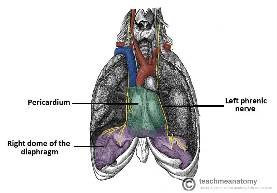
- Phrenic Nerve: Originates from the cervical spinal roots C3, C4, and C5; it provides motor and sensory innervation to the diaphragm.
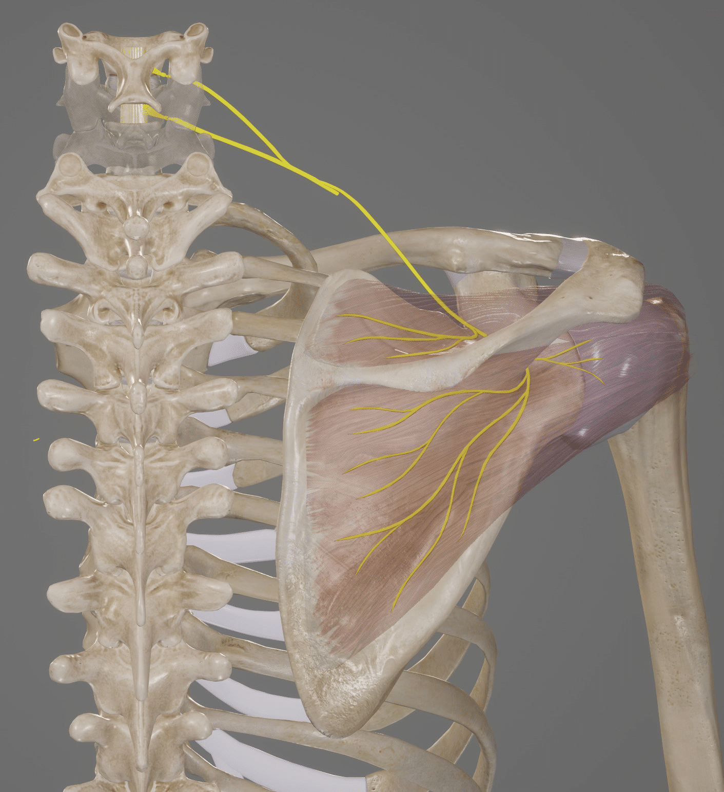
- Suprascapular Nerve: Supplies the supraspinatus and infraspinatus muscles.
:background_color(FFFFFF):format(jpeg)/images/article/the-hypoglossal-nerve/mUtVSxYk5qA79nKHFKQEA_course-of-hypoglossal-nerve_english.jpg)
:watermark(/images/watermark_5000_10percent.png,0,0,0):watermark(/images/logo_url.png,-10,-10,0):format(jpeg)/images/overview_image/2158/DDji56ULUN9e1nRlblXBw_triangles-neck-neurovasculature_english.jpg)
Arteries
- External Carotid Artery (ECA): It supplies blood to the face and neck.
- Internal Carotid Artery (ICA): Supplies blood to the brain.
- Vertebral Artery (VA): Supplies part of the brain, spinal cord, and neck muscles.
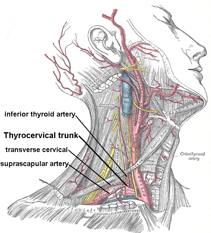
- Thyrocervical Trunk (TCT): Provides several branches that supply the neck and thyroid gland, including the inferior thyroid artery (ITA).
- Axillary Artery: Continuation of the subclavian artery, supplying the upper limb.


:background_color(FFFFFF):format(jpeg)/images/article/vertebral-artery/Pr3DUiOydFe8hJ3WXcIg_qZw8tXTIX5JHJ79oMmfKA_Vertebral_artery.png)

Veins
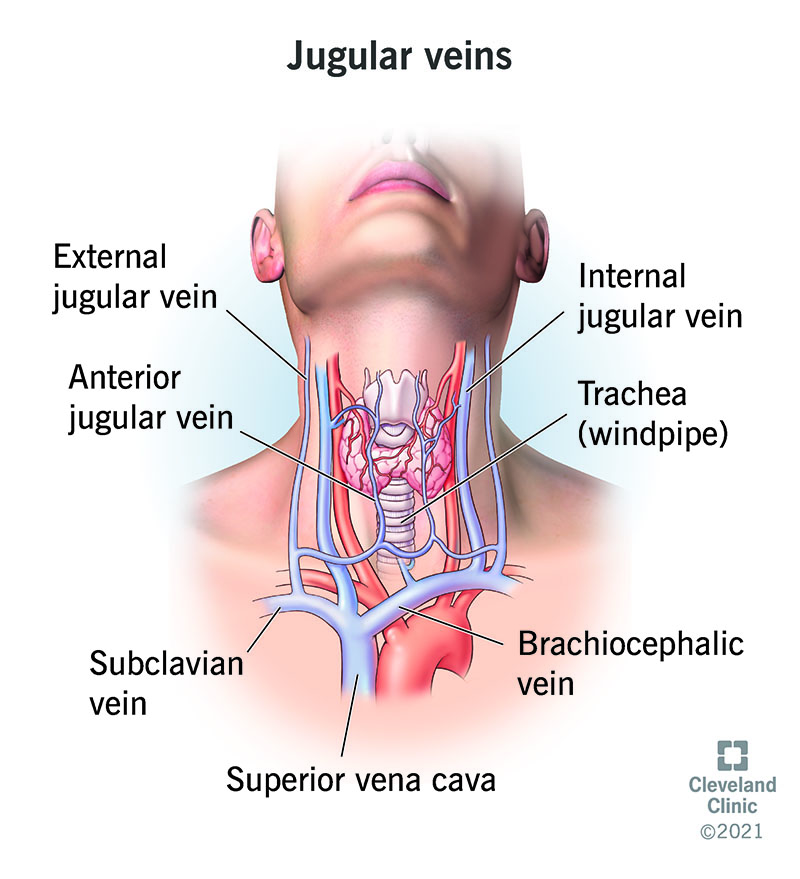
- Internal Jugular Vein: Drains blood from the brain, face, and neck.
- External Jugular Vein: Drains blood from the exterior of the cranium and deep parts of the face.
Brachial Plexus
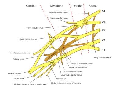
The brachial plexus is a network of nerve fibers that runs from the spine, through the neck, the axilla (armpit area), and into the arm. It consists of roots, trunks, divisions, cords, and terminal branches. - Roots: The starting point from spinal nerves C5 to T1. - Trunks: Formed by the combination of roots; they are named upper, middle, and lower. - Long Thoracic Nerve: Branches off the plexus and innervates the serratus anterior muscle.
Other Key Structures
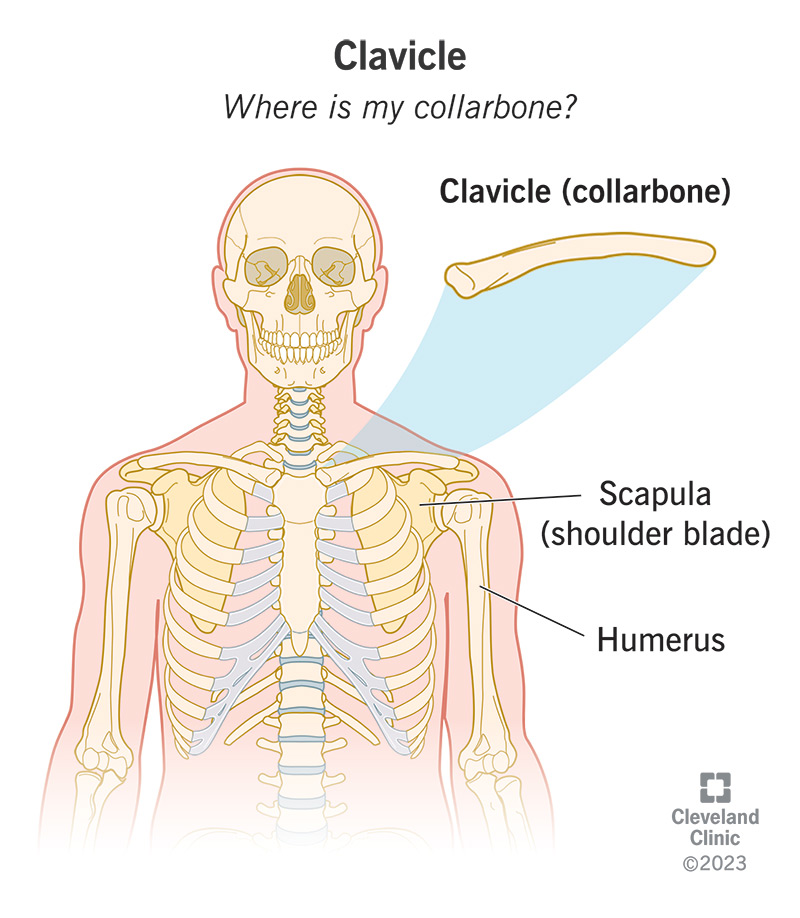
- Clavicle: Also known as the collarbone, it connects the arm to the body.
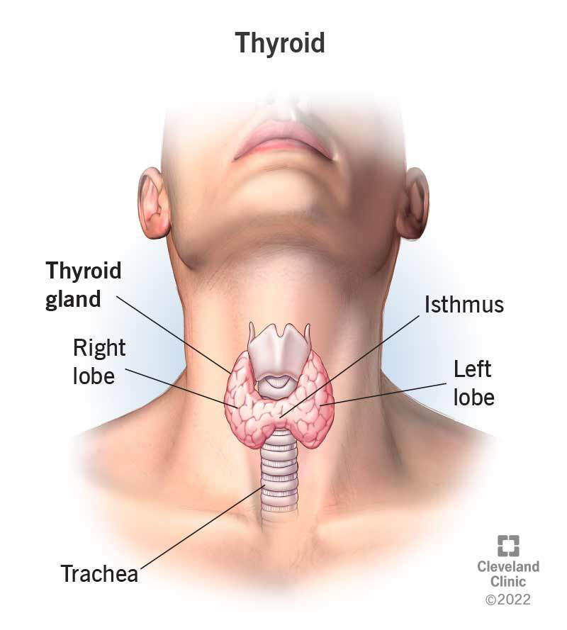
- Thyroid Gland: An endocrine gland producing hormones that regulate metabolism.
- Note from an M2: Make sure you know the nuances and differences between the different components of the thyroid gland.
- First Rib (Root of Neck): Provides an attachment point for muscles and supports the root of the neck.
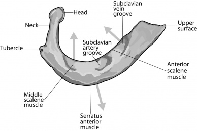
These anatomical landmarks play significant roles in the structural integrity, movement, and function of the neck and related structures, making their understanding crucial for medical and health-related studies.