Anatomy of the Ear
Anatomy of the Ear
External Ear
The external ear comprises the auricle (or pinna) and the external acoustic meatus. These structures are innervated primarily by the auriculotemporal branch of the mandibular nerve [V3], the lesser occipital nerve (C2), the great auricular nerve (C2, C3), and the auricular branch of the vagus nerve [X]. The auricle has several landmarks including the helix, antihelix, concha, tragus, antitragus, lobule, cymba conchae, triangular fossa, and the intertragic incisure. 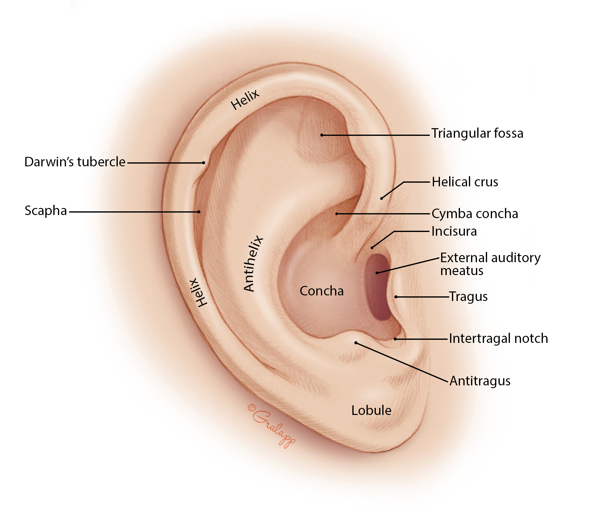
Middle Ear
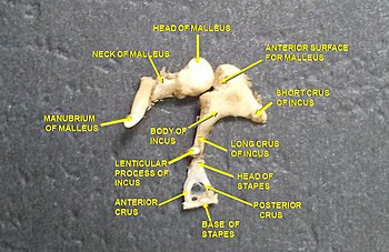
The middle ear, or tympanic cavity, contains the auditory ossicles: the malleus, incus, and stapes. The middle ear is connected to the pharynx via the pharyngotympanic tube (Eustachian tube) and to the internal ear via the oval window. The tympanic membrane separates the external ear from the middle ear and features notable landmarks such as the pars flaccida, pars tensa, cone of light, handle of malleus, and umbo.
Ossicles of the Middle Ear
- Malleus: The first of the ossicles, attached to the tympanic membrane.

- Incus: Located between the malleus and stapes.

- Stapes: The final ossicle, whose footplate attaches to the oval window, leading into the inner ear.
The ossicles transmit and amplify sound vibrations from the tympanic membrane to the internal ear. The tensor tympani muscle (innervated by CN V3) and the stapedius muscle (innervated by CN VII) help modulate these vibrations, especially during loud sounds, to protect the inner ear from damage. 
Walls of the Tympanic Cavity
- Tegmental wall (roof): Separates the tympanic cavity from the middle cranial fossa.
:watermark(/images/watermark_only_413.png,0,0,0):watermark(/images/logo_url_sm.png,-10,-10,0):format(jpeg)/images/anatomy_term/tegmental-wall-of-tympanic-cavity/jJ5qKi5qciIPmnB6m2NIIA_Tegmental_wall_of_tympanic_cavity.jpg)
- Jugular wall (floor): Separates the tympanic cavity from the internal jugular vein.
:watermark(/images/watermark_only_413.png,0,0,0):watermark(/images/logo_url_sm.png,-10,-10,0):format(jpeg)/images/anatomy_term/labyrinthine-wall-of-tympanic-cavity/62EQAeoBPZ3GqzTKPlQQ_Labyrinthine_wall_of_tympanic_cavity.jpg)
- Membranous (lateral) wall: Contains the tympanic membrane.
- Labyrinthine (medial) wall: Contains the oval window and the round window leading to the inner ear.
- Mastoid (posterior) wall: Contains openings to the mastoid air cells.
:watermark(/images/watermark_only_413.png,0,0,0):watermark(/images/logo_url_sm.png,-10,-10,0):format(jpeg)/images/anatomy_term/mastoid-wall-of-tympanic-cavity/vn61b6l9M4pikmut7OUOcw_Mastoid_wall_of_tympanic_cavity.jpg)
- Carotid (anterior) wall: Contains the opening of the pharyngotympanic (Eustachian) tube.
:watermark(/images/watermark_5000_10percent.png,0,0,0):watermark(/images/logo_url.png,-10,-10,0):format(jpeg)/images/overview_image/2902/UGbIHl7NS7p7ZEp7dIMQ_tympanic-cavity-wall_english.jpg)
Internal Ear
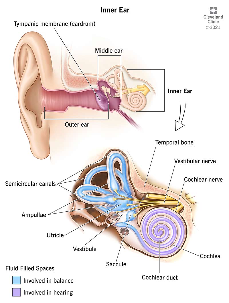
The internal ear consists of the cochlea, vestibule, and semicircular canals, all of which are housed within the bony labyrinth. The cochlea is responsible for hearing, while the vestibule and semicircular canals contribute to balance and spatial orientation. 
Nerve Supply
- Vestibulocochlear nerve (CN VIII): Supplies the cochlea (via the cochlear nerve) for hearing and the semicircular canals and vestibule (via the vestibular nerve) for balance.
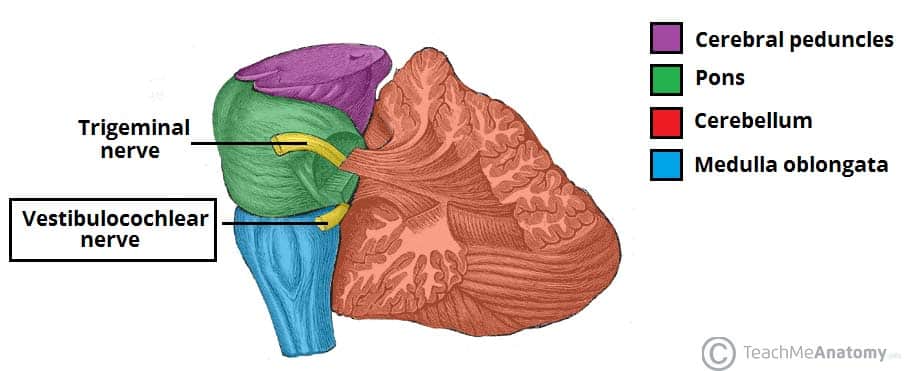
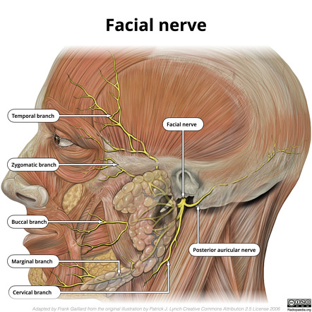
- Facial nerve (CN VII): Enters the internal acoustic meatus alongside the vestibulocochlear nerve and gives off several branches including the greater petrosal nerve (GPN), chorda tympani (CTM), and branches to the stapedius muscle.
Clinical Correlates
Tympanic Membrane Pathologies
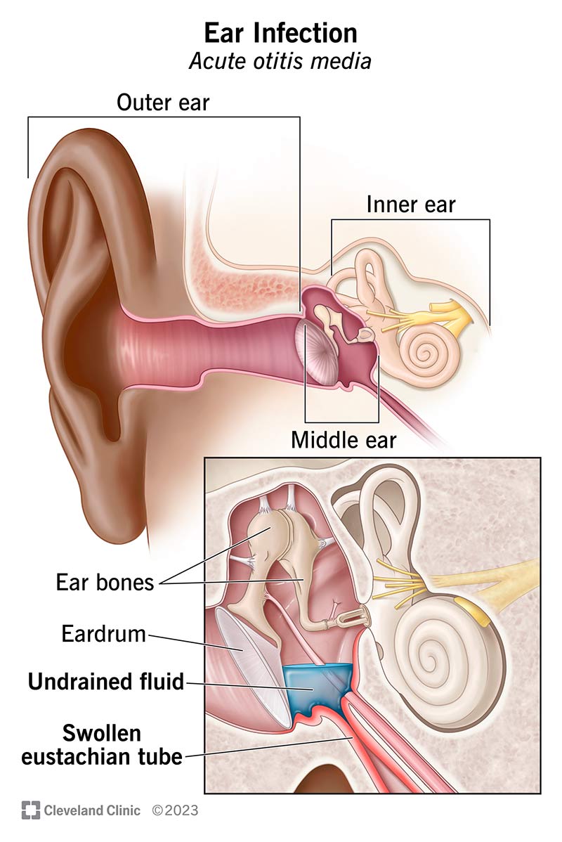
- Otitis Media: Infection of the middle ear, often resulting in a swollen and red eardrum.
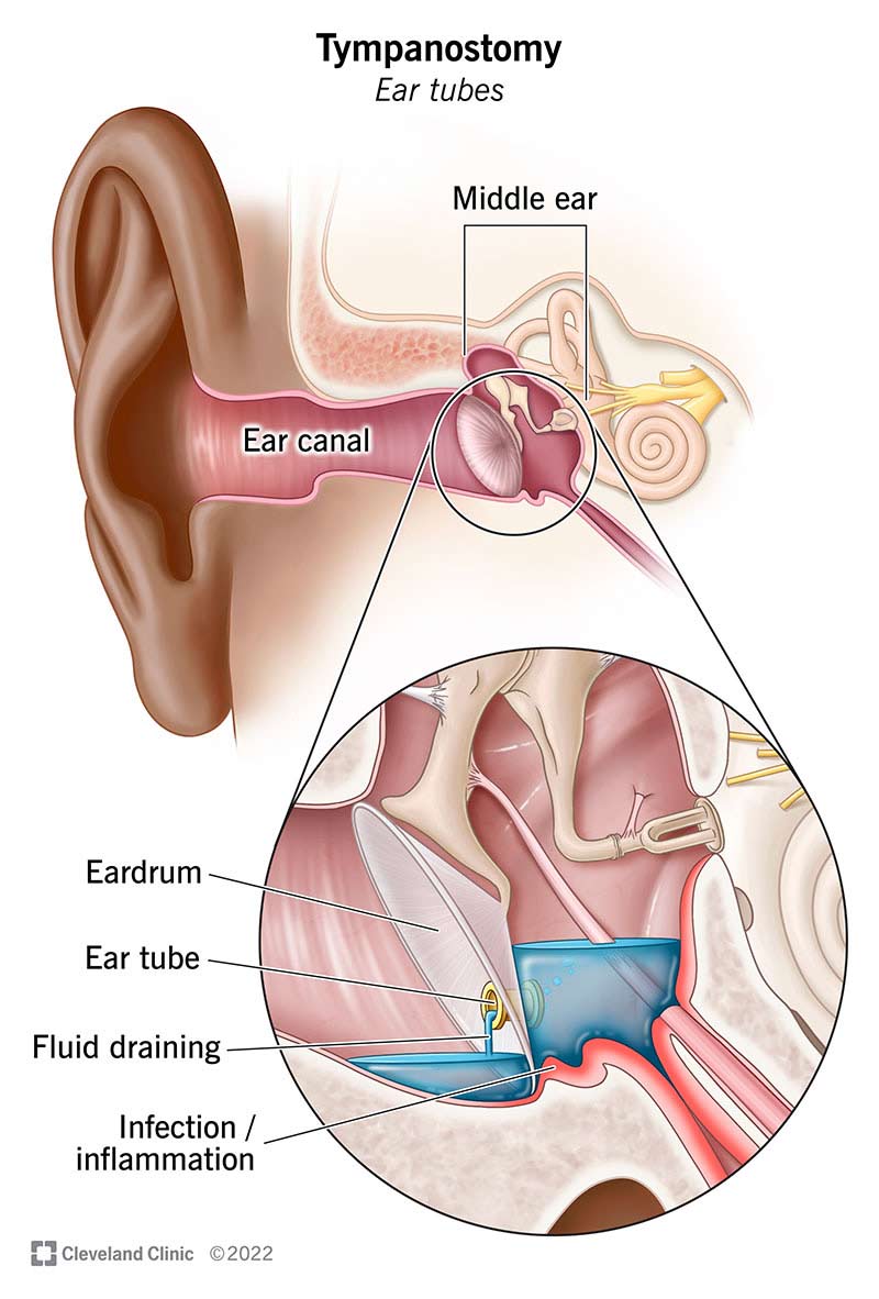
- Myringotomy Incision: A surgical incision in the tympanic membrane to relieve pressure or drain fluid.
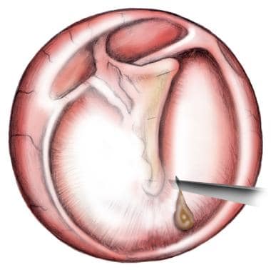
- Tympanostomy Tube Insertion: Placement of a tube in the tympanic membrane to allow continuous drainage and prevent fluid buildup.
Fracture and Disorders
- Fracture of the Tympanic Part of the Temporal Bone: Can lead to hearing loss and vestibular dysfunction due to damage to the internal ear structures.

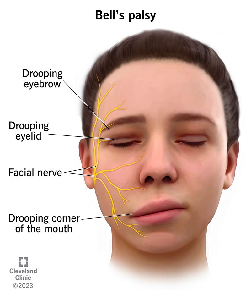
- Facial Nerve Paralysis: Damage to the facial nerve (CN VII) within the temporal bone can result in muscle paralysis on the affected side of the face.
