Nasal Cavity and Paranasal Sinuses
Nasal Cavity and Paranasal Sinuses
Nasal Cavity
Anatomy
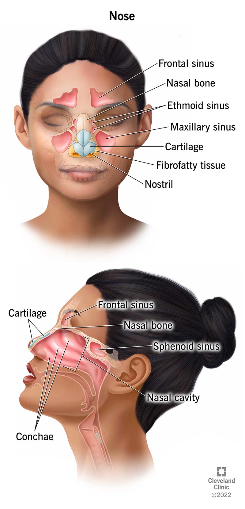
Roof: Formed by the nasal bone, frontal bone, cribriform plate of the ethmoid bone, and body of the sphenoid bone.
Floor: Composed of the palatine process of the maxilla and the horizontal plate of the palatine bone.
Medial Wall: The nasal septum, including the perpendicular plate of the ethmoid bone, vomer, and septal cartilage.
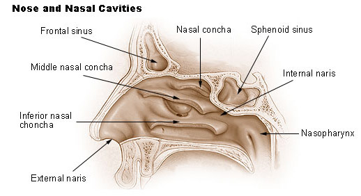
Lateral Wall: Consists of the superior, middle, and inferior conchae; the superior and middle conchae are parts of the ethmoid bone, while the inferior concha is a separate bone.

Relation to Surrounding Structures
Face: The anterior opening of the nasal cavity (nostrils) opens to the face.
Cranial Cavity: The nasal cavity is related to the anterior cranial fossa through the cribriform plate.
Orbit: The orbit is situated laterally and superiorly, bounded by the ethmoid bone.
Ethmoidal Cells: Located superiorly within the ethmoid bone, open into the middle meatus of the nasal cavity.
Maxillary Sinus: The lateral wall of the nasal cavity is related to the medial wall of the maxillary sinus.
Oral Cavity: The floor of the nasal cavity is shared with the roof of the oral cavity.
Surface Features
Conchae: Three bony projections (superior, middle, and inferior) increase surface area for air filtration and humidification.
Meatuses: Spaces below each concha (superior, middle, and inferior) where paranasal sinuses and nasolacrimal duct open.
Olfactory Region: Located in the upper part of the nasal cavity where olfactory receptors reside.
Paranasal Sinuses
Openings Within the Nasal Cavity
Frontal Sinus: Opens into the middle meatus via the frontonasal duct.
Maxillary Sinus: Opens into the middle meatus via the maxillary hiatus.
Ethmoidal Sinuses: Anterior, middle, and posterior groups open into the middle and superior meatuses.
Sphenoidal Sinus: Opens into the spheno-ethmoidal recess.
Nasolacrimal Duct: Drains tears from the lacrimal sac into the inferior meatus.

Blood Supply
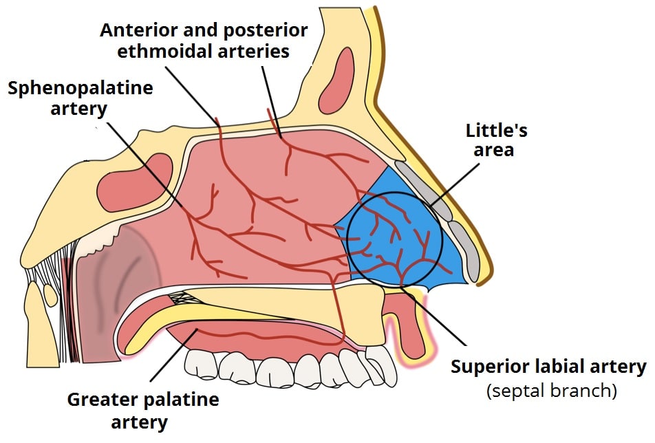
Ophthalmic Artery:
- Anterior Ethmoidal Artery: enters the nasal cavity through the anterior ethmoidal foramen.
- Posterior Ethmoidal Artery: enters through the posterior ethmoidal foramen.
Maxillary Artery:
- Sphenopalatine Artery: accompanies the nasopalatine nerve.
Facial Artery:
- Lateral Nasal Branches.

Key Anastomoses
The area where branches of the sphenopalatine, greater palatine, anterior ethmoidal, and superior labial arteries converge is prone to epistaxis (nosebleeds), notably Little's area.
Nerve Supply
Olfactory Nerve (CNI): Provides olfactory sensation.
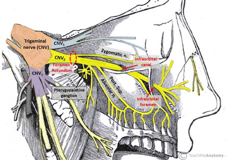
Trigeminal Nerve (CNV):
- V1 (Ophthalmic): Anterior ethmoidal nerve supplies the anterior and superior parts of the nasal cavity.
- V2 (Maxillary): Gives rise to the nasopalatine nerve and posterior nasal branches which supply the posterior and inferior parts.
Pterygopalatine Fossa
Contents and Fibers
Nerves:
- Zygomaticotemporal Nerve
- Zygomaticofacial Nerve
- Pharyngeal Nerve
- Nasal Nerves
- Orbital Branches, Infra-orbital Nerve, Posterior Superior Alveolar Nerve
- Greater and Lesser Palatine Nerves
- Pterygopalatine Ganglion
- Posterior Superior and Middle Superior Alveolar Nerves
- Pharyngeal Nerves
- Maxillary Nerve [V2]: Purely sensory, carries parasympathetic and sympathetic fibers to targets like the lacrimal gland, nasal mucosa, and upper salivary glands.
- Zygomatic Nerve, Posterior Superior Alveolar Nerve, 2 ganglionic branches
- Nerve of Pterygoid Canal: Formed by the union of greater petrosal nerve and deep petrosal nerve, contains both preganglionic parasympathetic (VII) and postganglionic sympathetic fibers (SCG and carotid plexus).

Pathways
Lacrimal Gland: Fibers travel with branches of V2, particularly the zygomaticotemporal nerve.
Other Glands: Fibers distribute to nasal mucosa and upper salivary glands via pterygopalatine ganglion, joining ganglionic branches of V2 to supply areas like the orbital wall, sphenoidal and ethmoidal sinuses, mucosa of the hard palate and adjacent gingiva, lateral and medial walls of the nasal cavity, and glands of the nasopharynx.