Interior Cranium, Meninges, and Orbit
Lecture Objectives Part 1
- Identify the bones of the interior of the skull.
- Demonstrate the bony landmarks of the interior of the skull.
- Describe the boundaries of the cranial fossae (anterior, middle, posterior).
- List the contents of the cranial fossae.
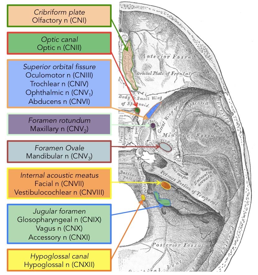
- Identify the exit points of all twelve cranial nerves in the cranial cavity.
- Identify the bones and major openings at the exterior base of the skull.

Sutures and Osteology
- Sutures: Not closed at birth, with cartilage component; fuse between ages 16-24.
- Sutures to review: Sphenosquamous suture, Coronal suture, Squamous suture, Sphenoparietal suture.
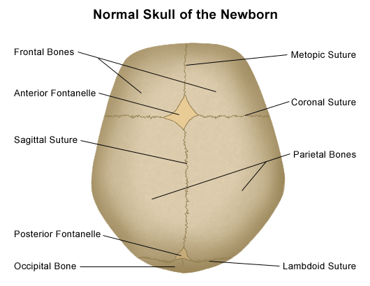
Bony Landmarks:
- Frontal Bone: Superciliary arch, Supra-orbital notch, Glabella, Nasion
- Parietal Bone: Pterion, Sphenoparietal suture
- Occipital Bone: External occipital protuberance, Lambda
- Temporal Bone: Squamous part, Zygomatic process, Mastoid process, Styloid process, Mandibular fossa

Cranial Fossae Boundaries and Contents:
Anterior Cranial Fossa:
- Bones: Frontal, Ethmoid, Sphenoid (lesser wing)
- Notable Structures: Crista galli, Cribriform plate, Foramen cecum
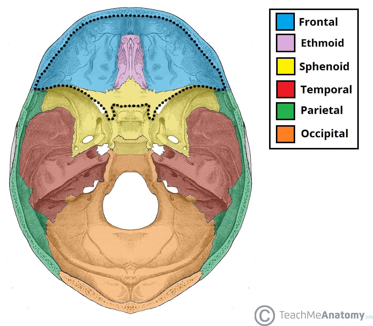
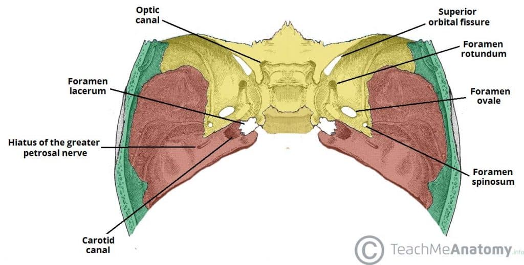
Middle Cranial Fossa:
- Bones: Sphenoid (greater wing, body), Temporal (squamous, petrous parts)
- Notable Structures: Sella turcica (Tuberculum sellae, Hypophysial fossa, Dorsum sellae), Superior orbital fissure, Foramen rotundum, Foramen ovale, Foramen spinosum, Carotid canal, Groove for the middle meningeal artery
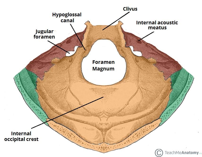
Posterior Cranial Fossa:
- Bones: Occipital, Temporal (mastoid part), Sphenoid (posterior body)
- Notable Structures: Foramen magnum, Internal acoustic meatus, Jugular foramen, Hypoglossal canal, Clivus, Internal occipital protuberance
Exit Points of Cranial Nerves:
- Cribriform plate: CN I (Olfactory nerve)
- Optic canal: CN II (Optic nerve)
- Superior orbital fissure: CN III (Oculomotor nerve), CN IV (Trochlear nerve), CN V1 (Ophthalmic division of Trigeminal nerve), CN VI (Abducent nerve)
- Foramen rotundum: CN V2 (Maxillary division of Trigeminal nerve)
- Foramen ovale: CN V3 (Mandibular division of Trigeminal nerve)
- Internal acoustic meatus: CN VII (Facial nerve), CN VIII (Vestibulocochlear nerve)
- Jugular foramen: CN IX (Glossopharyngeal nerve), CN X (Vagus nerve), CN XI (Accessory nerve)
- Hypoglossal canal: CN XII (Hypoglossal nerve)
Lecture Objectives Part 2 (Meninges):
- Describe the specializations of the cranial meninges and the dural venous sinuses related to them.
- Trace a drop of venous blood through the dural venous sinuses.
- Point out the course and distribution of the middle meningeal artery.
- Describe the cavernous sinus and the relationship of its contents with each other.
- Explain the concept of an “emissary vein” and place it into clinical context.

Cranial Meninges:
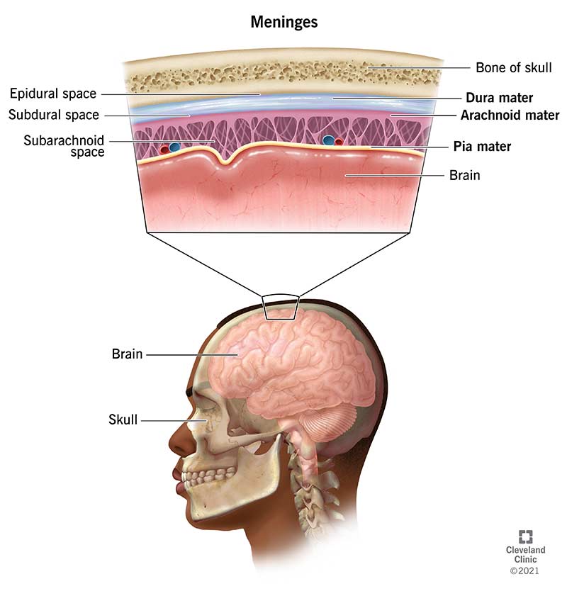
The meninges consist of three layers: - Dura Mater (periosteal and meningeal layers): Thick, durable membrane closest to the skull. - Arachnoid Mater: Middle layer with web-like extensions that bridge the subarachnoid space. - Pia Mater: Innermost delicate layer that adheres tightly to the surface of the brain.
Dural Septa and Sinuses:
- Falx cerebri: Separates the two cerebral hemispheres.
- Tentorium cerebelli: Separates the cerebrum from the cerebellum.
- Falx cerebelli: Separate two hemispheres of the cerebellum.
- Diaphragma sellae: Small septum between the hypothalamus and pituitary gland.
The venous sinuses, which include the Superior sagittal sinus, Inferior sagittal sinus, Straight sinus, and Transverse sinuses, drain venous blood from the brain.
Vascular Supply and Hematomas:
Arterial Supply:
- Middle meningeal artery: Major supply to the meninges, branch of the maxillary artery.
Hematomas:
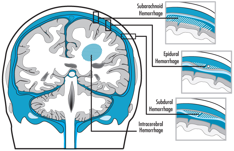
- Epidural Hematoma: Accumulation of blood between the dura mater and the skull, commonly associated with arterial ruptures, particularly the middle meningeal artery.
- Subdural Hematoma: Accumulation of blood between the dura and arachnoid mater, typically venous, resulting from the shearing of bridging veins.
Dural Venous Sinuses:
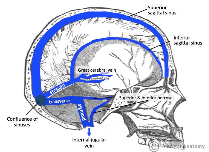
- Superior sagittal sinus: Located along the attached margin of the falx cerebri.
- Inferior sagittal sinus: Located in the inferior margin of the falx cerebri.
- Straight sinus: Connects the inferior sagittal sinus to the confluence of sinuses.
- Transverse sinuses: Paired sinuses that extend laterally from the confluence of sinuses.
- Sigmoid sinuses: Continuations of the transverse sinuses that lead to the jugular foramen.
- Cavernous sinus: Located on each side of the sella turcica, receiving ophthalmic veins, and communicating with the pterygoid plexus and superior and inferior petrosal sinuses.
Lecture Objectives Part 3 (Orbit):
- Identify the bones that form the walls and margins of the orbits.
- Describe the arteries and veins of the orbit.
- List the nerves contained within the orbit and state the function of each.
- Describe the structure, function, and innervation of the extraocular muscles.
- Identify and name the surface parts of the eyeball and the eyelids.
- Trace the path of a tear from the lacrimal gland through the lacrimal apparatus, naming all of the structures that it traverses.
- Understand how the autonomic nervous system innervates and controls the sphincter and dilator pupillae muscles, the ciliary body of the eye, and the levator palpebrae superioris muscle of the eyelid.

Bony Orbit:
- Shape/Orientation: Pyramidal structure with four walls (roof, floor, medial wall, lateral wall) and an apex.
- Bones: Frontal bone, Zygomatic bone, Maxilla, Palatine bone, Lacrimal bone, Ethmoid bone, Sphenoid bone.
Eyelid Structure:
- Orbital Septum and Tarsal Plates: Connective tissue layers that protect and provide structure to the eyelids.

Fascial Specializations:
- Periorbita: Periosteum lining the orbit.
- Fascial Sheath: Encloses the eyeball, forming the socket.
- Suspensory Ligament: Supports the eyeball within the orbit.
- Check Ligaments: Restrict excess movement of the eyeball.
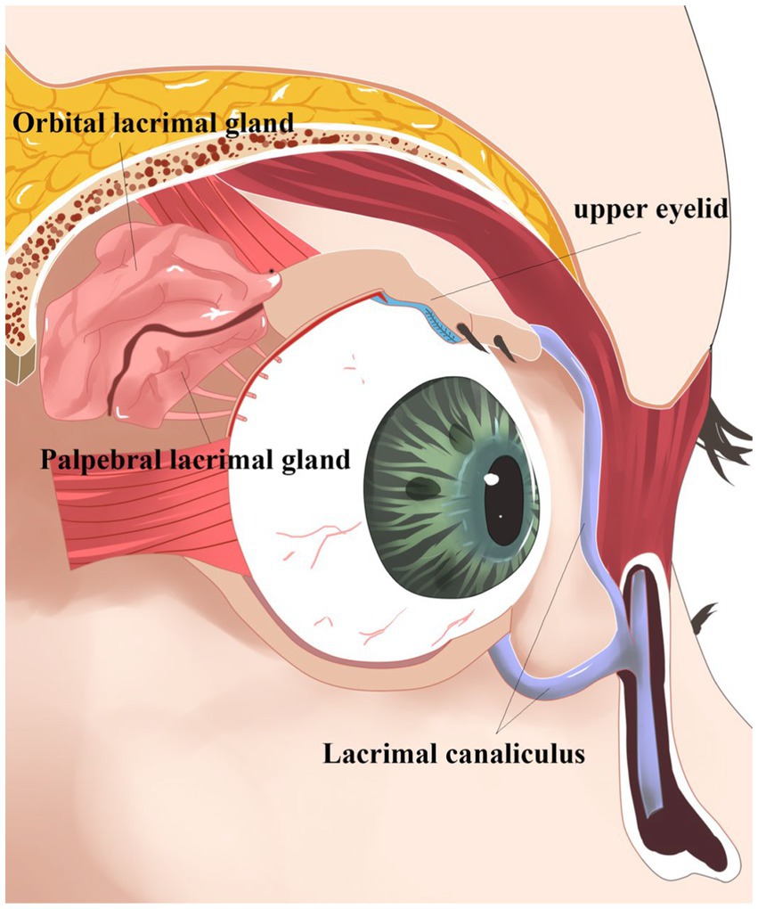
Lacrimal Apparatus:
- Path of a Tear: Lacrimal gland → Lacrimal puncta → Lacrimal canaliculi → Lacrimal sac → Nasolacrimal duct.
Vessels of the Orbit:
- Arteries: Ophthalmic artery (main supply), Lacrimal artery, Supra-orbital artery, Ethmoidal arteries.
- Veins: Superior and Inferior ophthalmic veins, Angular vein, Pterygoid plexus of veins.
:background_color(FFFFFF):format(jpeg)/images/article/arteries-and-veins-of-the-orbit/DTaCrffG5M6KqpZz8n0u6g_Blood_vessels_of_orbit_-_superior_view.png)
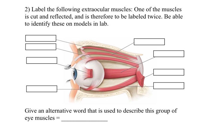
Extraocular Muscles:
- Six Extraocular Muscles: Superior rectus, Inferior rectus, Medial rectus, Lateral rectus, Superior oblique, Inferior oblique.
- Innervation: CN III (Oculomotor nerve), CN IV (Trochlear nerve), CN VI (Abducent nerve).
Nerve Supply Mnemonic:
- LR6: Lateral Rectus is innervated by CN VI (Abducent nerve).
- SO4: Superior Oblique is innervated by CN IV (Trochlear nerve).
- All the rest are innervated by CN III (Oculomoto...