Bones and Superficial Structures of the Neck
Bones and Superficial Structures of the Neck
Surface Anatomy of the Neck
Key landmarks and structures: Frontal bone, Zygomatic bone, Supraorbital margin, Infraorbital margin, Helix, Philtrum, Antihelix, Commissure of lips, Tragus, Antitragus, Mental protuberance, Mandibular angle, Submandibular gland, Mandible (inferior border), Thyroid cartilage (Adam's apple), Trapezius muscle, Clavicle, Suprasternal notch, Jugular notch, Sternocleidomastoid muscle (innervated by accessory CN).

Hyoid Bone
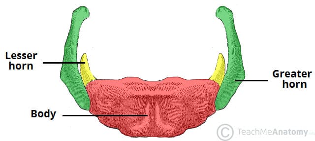
Location: Anteriorly located at the junction of the soft structures of the floor of the mouth and the larynx, at C3 vertebra level.
Parts: Body, Greater horn (palpable laterally), Lesser horn.
Cartilages of the Neck
Thyroid Cartilage: Forms the laryngeal prominence (Adam's apple) and is located 1-2 cm below the hyoid bone.
Cricoid Cartilage: Located just inferior to the thyroid cartilage at C6 vertebra level. Forms a complete ring.

Cartilaginous Ring of Trachea: Site of emergency cricothyrotomy.

Organization of the Neck
Inferior Border: Sternocleidomastoid muscle.
Fascia: Divided into investing layer, pretracheal fascia (muscular and visceral layers), and prevertebral fascia. The pretracheal fascia extends from the hyoid bone to the pericardium and encloses the infrahyoid muscles, thyroid, trachea, larynx, pharynx, and esophagus.
Layers of Fascia of the Neck
Superficial Fascia: Contains platysma muscle (aids in facial expression, relieves pressure from superficial veins when contracted).
Deep Fascia: Investing layer (prevents spread of infection), Pretracheal layer (surrounds infrahyoid muscles, thyroid, trachea, larynx), Prevertebral layer (surrounds vertebral column, extends laterally as axillary sheath).
Superficial Veins of the Neck
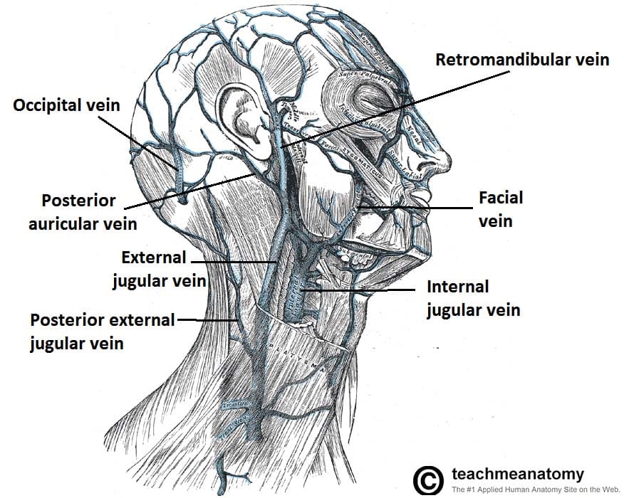
Major Veins: External jugular vein, Anterior jugular vein, Posterior auricular vein.
Drainage: External jugular vein drains into the subclavian vein.
Cutaneous Branches of the Cervical Plexus
Nerves: Lesser Occipital n. (C2), Greater Auricular n. (C2 and C3), Transverse Cervical n. (C2 and C3), Supraclavicular nn.
Muscles and Contents of the Posterior Triangle of the Neck
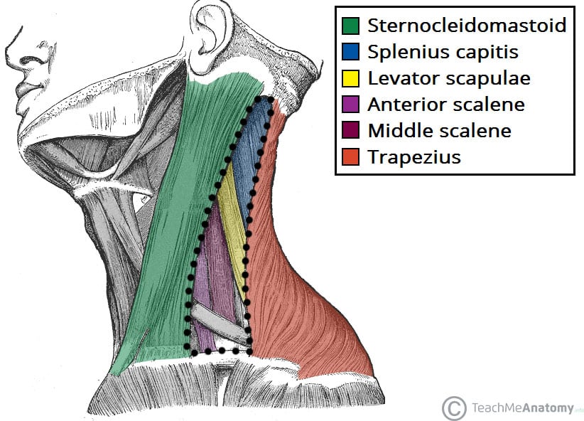
Borders: Defined by the sternocleidomastoid, trapezius, and clavicle.
Muscles: Splenius capitis, Levator scapulae, Scalene muscles (anterior, middle, posterior), Omohyoid.
Nerves: Cutaneous branches exit at Erb’s Point, CN XI (Accessory nerve).
Vessels: Superficial and deep vessels.
Sternocleidomastoid (SCM) Muscle
Attachments: Sternum, clavicle, mastoid process.
Actions: Bilateral contraction causes neck flexion; unilateral contraction causes head rotation and lateral flexion.
:background_color(FFFFFF):format(jpeg)/images/article/sternocleidomastoid-muscle/N21GRSiWKOf8BQlrcQhA_hbimApe3IQfTimfNbNgnBw_sternocleido_mastoid.png)
Scalene Muscles
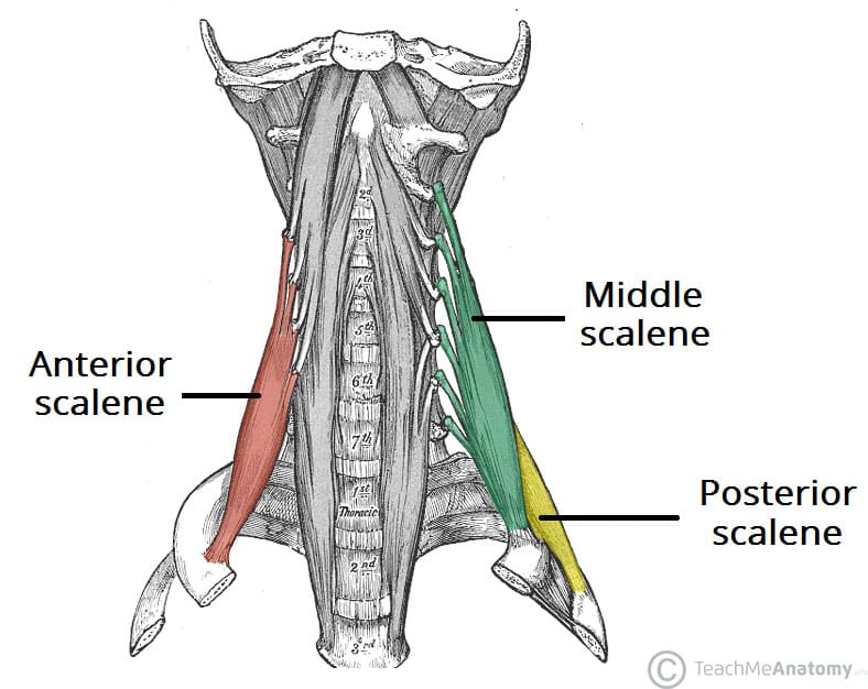
Attachments: Transverse processes of cervical vertebrae to first and second ribs.
Actions: Elevate ribs, assist in neck flexion and lateral flexion.
Relations: Subclavian artery and brachial plexus pass between anterior and middle scalene muscles.
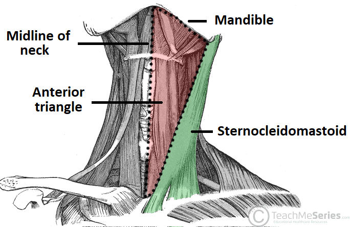
Anterior Cervical Triangle
Subdivisions: Submandibular Triangle, Carotid Triangle, Muscular Triangle.
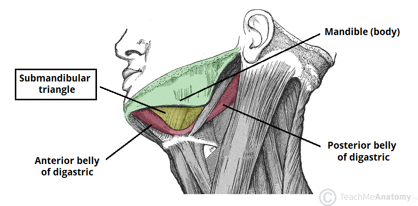
Submandibular Triangle
Contents: Submandibular gland, facial artery, and vein.
Carotid Triangle
Contents: Carotid arteries, internal jugular vein, vagus nerve (CN X), deep cervical lymph nodes.
Muscular Triangle
Contents: Infrahyoid muscles (omohyoid, sternohyoid, sternothyroid, thyrohyoid), thyroid gland, larynx, trachea.
Suprahyoid and Infrahyoid Muscles
Suprahyoid Muscles: Digastric (anterior and posterior bellies), Stylohyoid, Mylohyoid, Geniohyoid. Functions primarily to elevate the hyoid bone and assist in opening the mandible.
Infrahyoid Muscles: Omohyoid, Sternohyoid, Sternothyroid, Thyrohyoid. Functions to depress the hyoid bone and larynx during swallowing and speaking.

Nerves of the Neck
CN X (Vagus Nerve): Major nerve of the carotid triangle with branches including superior and recurrent laryngeal nerves.
CN XI (Accessory Nerve): Innervates SCM and trapezius muscles, courses through the posterior triangle.
CN XII (Hypoglossal Nerve): Supplies most muscles of the tongue, travels within the submandibular triangle.
Ansa Cervicalis: Provides motor innervation to most of the infrahyoid muscles.
Phrenic Nerve (C3-C5): Innervates the diaphragm, important for breathing.
