Human Structure: Review of Unit 1
Skin, Superficial Back Muscles, Vertebral Canal, and Spinal Cord
Integumentary System
- Functions: Protection, containment, prevention of dehydration, heat regulation, sensation, synthesis, and storage of vitamin D.
- Structure:
- Epidermis: Protective barrier.
- Dermis: Contains nerve endings, sweat glands, oil glands, and hair follicles.
- Hypodermis (Subcutaneous): Fat and connective tissue.

- Components:
- Hair shaft, root, and follicle
- Sebaceous glands: Lubricate skin
- Sweat glands: Eccrine and apocrine
- Sensory nerve fibers: Meissner's corpuscle, Pacinian corpuscle
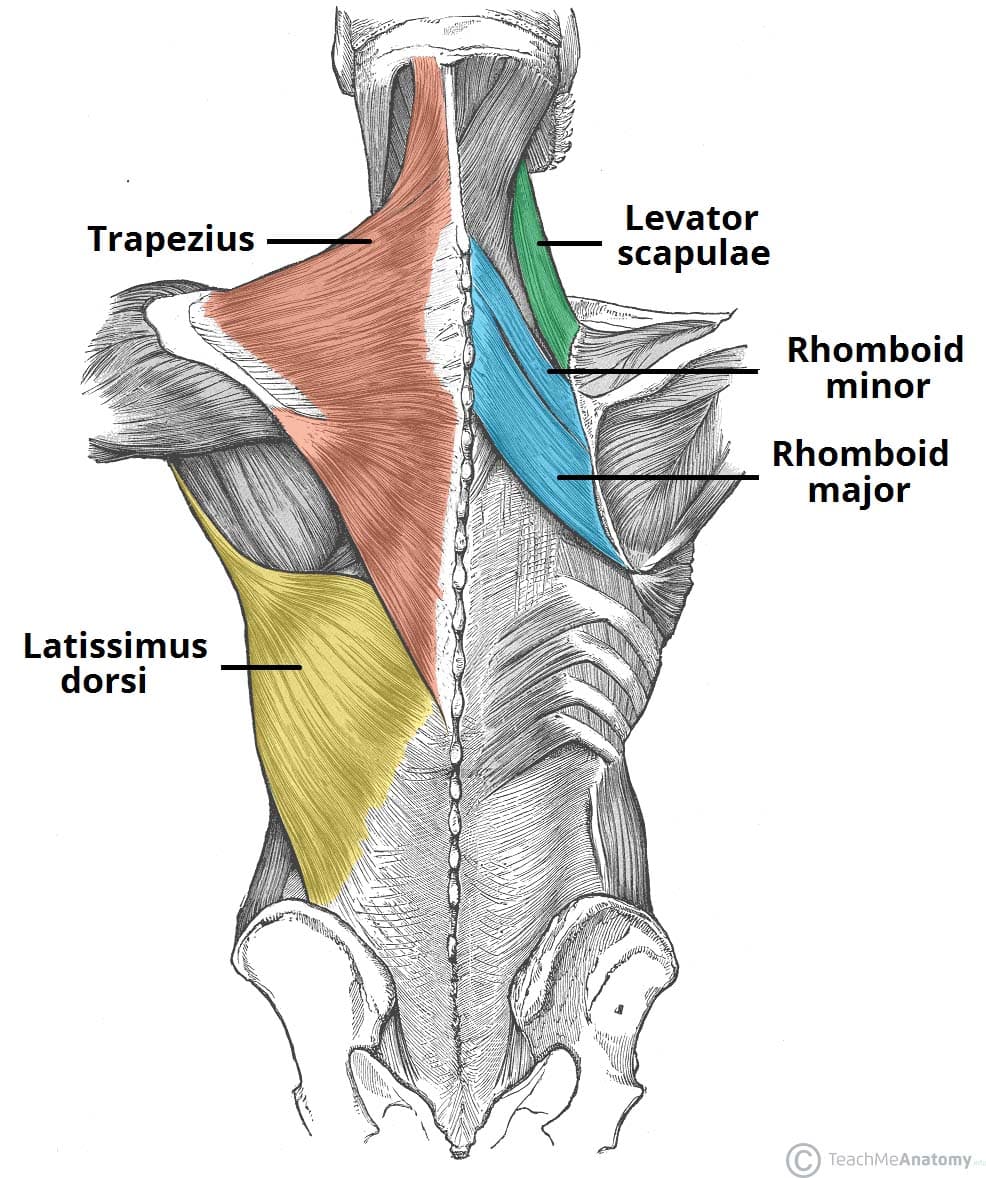
Superficial Back Muscles
Trapezius Muscle
- Innervation: Accessory nerve (CN XI) and ventral rami of C3 and C4
- Blood Supply: Transverse cervical artery
Latissimus Dorsi Muscle
- Innervation: Thoracodorsal nerve (posterior cord of brachial plexus)
- Blood Supply: Thoracodorsal artery
- Insertion: Floor of the intertubercular sulcus of humerus
:watermark(/images/watermark_5000_10percent.png,0,0,0):watermark(/images/logo_url.png,-10,-10,0):format(jpeg)/images/overview_image/942/C5TmNQeZx2RAJMPiDQiQ_Superficial-muscles-of-the-back_english.jpg)
Other Back Muscles
- Levator Scapulae
- Rhomboid Major
- Rhomboid Minor
Vertebral Canal and Spinal Cord
Sympathetic Nervous System
- T4 Sympathetic Ganglion
Spinal Nerve Components
- Dorsal Root: Sensory or afferent
- Ventral Root: Motor or efferent
- Rami Communicantes: Connect spinal nerves to the sympathetic trunk
- White and gray rami communicantes: Lateral in thoracic region, move anteriorly in the abdomen.
Dermatomes
- Areas of skin supplied by afferent nerve fibers from a single posterior spinal root.
- Key dermatomes: C4, C5, T1-T5, T7, T8.

Breast and Pectoral Muscles
Mammary Gland
- Structures: Areola, nipple, gland lobules, lactiferous ducts, and sinuses
- Functions: Split into 15-20 lobes, not encapsulated
Lymphatic Drainage
- Nodes: Subareolar plexus, circumareolar plexus, parasternal nodes
- Main Paths: Principal axillary lymph path, accessory axillary lymph path
Skeleton of the Pectoral Region
- Includes: Manubrium, sternum, scapula, clavicle, true ribs (1-7), costal cartilages
- Axial Skeleton: Head, neck, trunk
- Appendicular Skeleton: Shoulder girdle, pelvic girdle, extremities
Anterior Thoracic Wall
- Pectoralis Major Muscle
- Innervation: Medial and lateral pectoral nerve
- Blood Supply: Pectoral branch of the thoracoacromial trunk
- Pectoralis Minor Muscle
- Innervation: Medial pectoral nerve
Upper Appendicular Skeleton
- Structures: Clavicle, scapula, humerus

Back and Shoulder Muscles, Vertebral Canal, and Spinal Cord
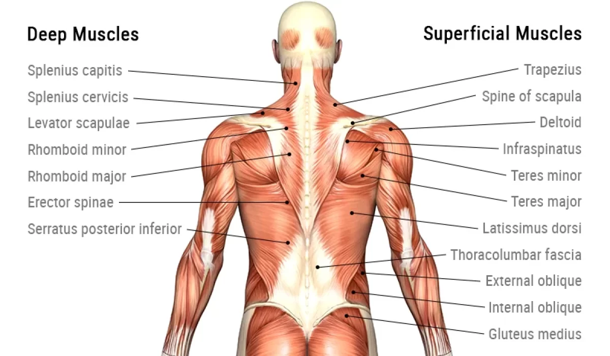
Superficial Back Muscles
- Layers:
- Superficial Layer: Trapezius, latissimus dorsi (extrinsic muscles)
- Intermediate Layer: Serratus posterior superior, inferior
- Deep Layer: Splenius, erector spinae, transversospinalis (intrinsic muscles)
Erector Spinae Muscles
- Muscles: Spinalis, longissimus, iliocostalis
- Innervation: Branches of the posterior rami
Spinal Cord and Nerve Roots
- Layers and Spaces:
- Epidural Space
- Dura Mater
- Arachnoid Mater
- Subarachnoid Space (with CSF)
- Pia Mater
Adult Spinal Cord
- Ends at: L1/L2 intervertebral disc
- Dural Sac: Ends at S2 vertebral level
- Filum Terminale Externum: Attaches to the dorsal coccyx
Upper Limb – Shoulder
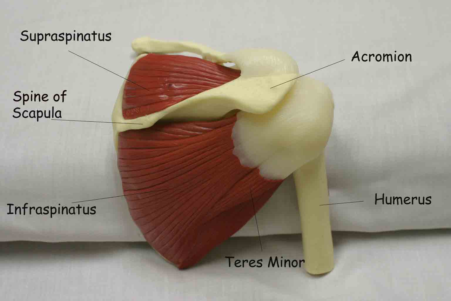
Rotator Cuff
- Muscles: Supraspinatus, infraspinatus, subscapularis, teres minor
- Function: Stabilizes the glenohumeral joint
Injury
- Supraspinatus injury: Prevents initiation of abduction
- Abduction of Upper Limb: Deltoid and supraspinatus muscles
Anatomy of the Arteries
Blood Supply to the Arm and Shoulder
- Subclavian Artery
- Branches: Thyrocervical trunk, suprascapular artery
- Axillary Artery
- Parts:
- Superior thoracic artery
- Thoracoacromial, lateral thoracic arteries
- Subscapular, anterior, posterior circumflex humeral arteries
- Parts:

Thoracic Wall, Pleural Cavity, and Lungs
Thoracic Wall
- Structures: Subclavian artery, vein, long thoracic nerve, lateral thoracic artery, serratus anterior muscle
Pleura and Pleural Cavity
- Serous Membrane:
- Visceral Pleura
- Parietal Pleura (cervical, costal, mediastinal, diaphragmatic parts)
Recesses and Reflections
- Terms: Costodiaphragmatic recess (important for fluid drainage), costomediastinal recess, sternal junction of costal and mediastinal pleurae, vertebral junction of costal and mediastinal pleurae

Hemothorax and Thoracentesis
- Hemothorax: Blood in the pleural cavity
- Thoracentesis: Procedure to remove fluid

Lymphatic Drainage of the Lungs
- Structures: Paratracheal nodes, bronchomediastinal lymphatic trunk, thoracic duct, bronchopulmonary nodes, pulmonary nodes
*You don't need to memorize the nodes
Pericardium and Heart
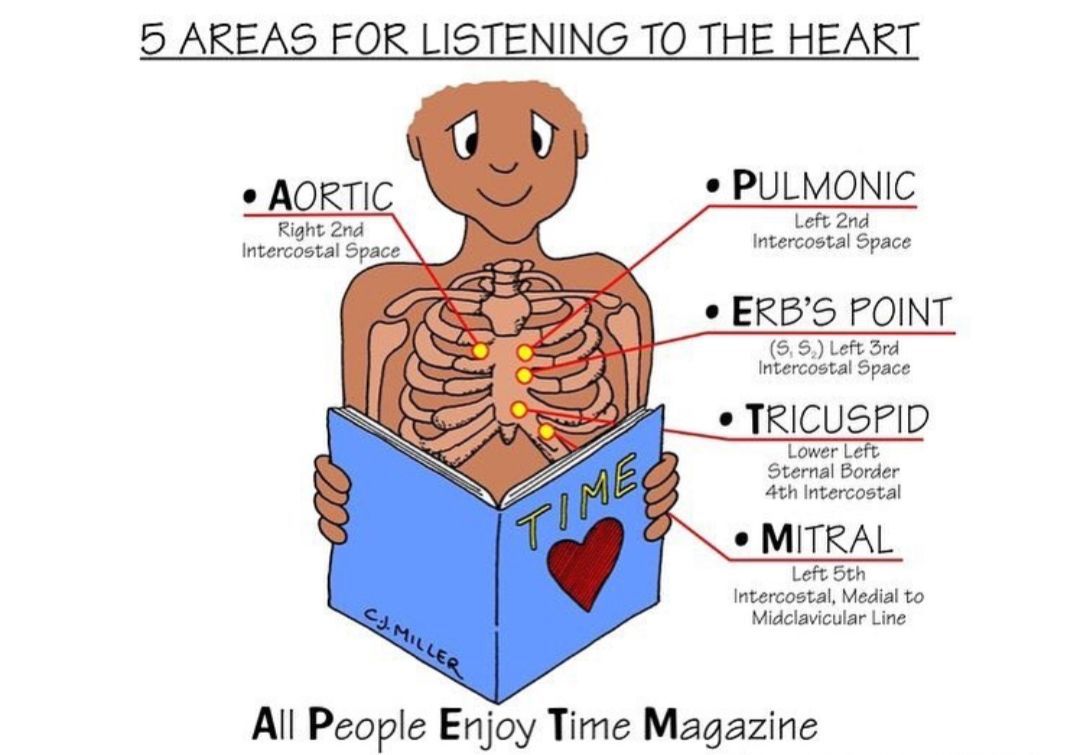
Auscultatory Areas
- Locations:
- 2nd Intercostal Space: Pulmonary, aortic sounds
- 5th Intercostal Space: Mitral, tricuspid sounds
Surface Projections of Heart Valves
- Site of Valves: PAMT (Pulmonary, Aortic, Mitral, Tricuspid)

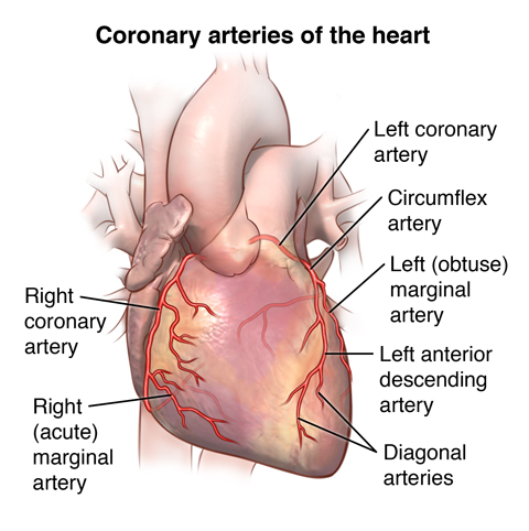
Coronary Arteries
- Arteries: Left coronary artery (LCA), right coronary artery (RCA)
- Branches: Circumflex, marginal branch, etc.
Conducting System of the Heart
- Components: SA node, AV node, Purkinje fibers
- Innervation:
- Sympathetic: Increases heart rate
- Parasympathetic (Vagus Nerve): Decreases heart rate
Mediastinum
Superior Mediastinum
- Contains: Thymus, great vessels, vagus & phrenic nerves, trachea, esophagus, thoracic duct
Posterior Mediastinum
- Structures: Esophagus, thoracic aorta, azygos vein, hemi-azygos vein, thoracic duct, vagus nerves, sympathetic trunk, splanchnic nerves


*They will test you on this content
Upper Limb Anatomy
Brachial Plexus
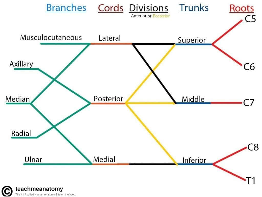
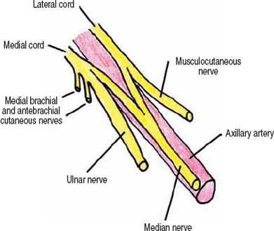
- Structure: 5 roots, 3 trunks, 3 anterior divisions, 3 posterior divisions, cords, terminal branches
- Mnemonic: Real Teachers Drink Cold Beer (Roots, Trunks, Divisions, Cords, Branches)
Important Nerves
- Nerves: Suprascapular, phrenic, dorsal scapular, long thoracic, etc.
Arm Muscles
Posterior Compartment
- Triceps Brachii (Long, Lateral, Medial heads): Infraglenoid tuberosity attachment, extends shoulder and elbow
- Anconeus: Extends elbow
Forearm and Hand
Flexor-Pronator Muscles
- Origin: Common tendon to medial epicondyle
- Muscles:
- Pronator Teres
- Flexor Carpi Radialis
- Palmaris Longus
- Flexor Carpi Ulnaris (ulnar nerve)
- Flexor Digitorum Superficialis (intermediate group)
- Brachioradialis: Flexor of forearm

Rotators of Radius
- Pronation: Supinator, pronator teres
- Supination: Supinator muscle

Brachial Artery
- Branches: Radial collateral, middle collateral, superior ulna collateral artery
Hand Anatomy
Carpal Tunnel
- Components: Carpal bones, flexor retinaculum
- Contents: Flexor tendons, synovial sheaths, median nerve
- Syndrome: Compression causing diminished sensation, thumb weakness
Movements of the Hand
- Thumb: Flexion, extension, opposition
Innervation of Hand Muscles
- Median Nerve: LOAF muscles (Lateral 2 lumbricals, Opponens Pollicis, Abductor Pollicis Brevis, Flexor Pollicis Brevis)
- Ulnar Nerve: All other intrinsic muscles

Cutaneous Innervation
- Notable Innervations: Lateral cutaneous nerve of forearm, medial cutaneous nerve of forearm, branches of radial and ulnar nerves
Here's a song to review:
