Upper Extremity Anatomy and Clinical Correlations
Upper Extremity Anatomy and Clinical Correlations

Shoulder Girdle
Bones
The shoulder girdle is composed of four primary bones: the clavicle, scapula, humerus, and the joints that connect them.
[h3]skeletal structure of shoulder[/h3]Joints
- Sternoclavicular Joint: This is a synovial joint located at the medial end of the clavicle. It is the only attachment of the upper limb to the axial skeleton. Despite being strong, it is quite mobile. Stability is provided by ligaments such as the costoclavicular ligament, the sternoclavicular ligament, and the interclavicular ligament. Injuries to this joint can lead to arthritis (osteoarthritis, rheumatoid arthritis, septic arthritis) and dislocations.
- Acromioclavicular Joint: This joint includes the acromion of the scapula and the clavicle. Injuries and arthritis can also affect this joint.
- Glenohumeral Joint: It is a synovial ball and socket joint, the most mobile joint in the human body but also the most prone to dislocation. Anterior dislocations are more common and less serious compared to posterior dislocations.
- Scapulothoracic Joint: A functional joint formed by the articulation between the anterior surface of the scapula and the posterior thoracic rib cage.
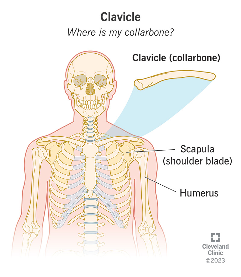
Clavicle
The clavicle is a long bone that transmits force from the upper limb to the axial skeleton. Clavicular fractures can occur due to falls onto the shoulder or outstretched hands. These fractures are arbitrarily divided into thirds: lateral (15%), middle (80%), and medial (5%). After a fracture, the lateral end of the clavicle is displaced inferiorly by the weight of the arm, while the medial end is pulled superiorly by the sternocleidomastoid muscle.
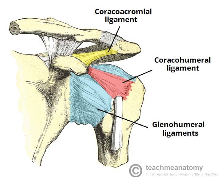
Glenohumeral Joint
This synovial ball and socket joint is the most mobile and consequently the most unstable joint in the human body. Shoulder dislocations, especially anterior dislocations (95%), are common in adolescents and young adults, often as athletic injuries. The axillary nerve may be injured. Posterior shoulder dislocations are less common and typically result from electrocution or seizure.
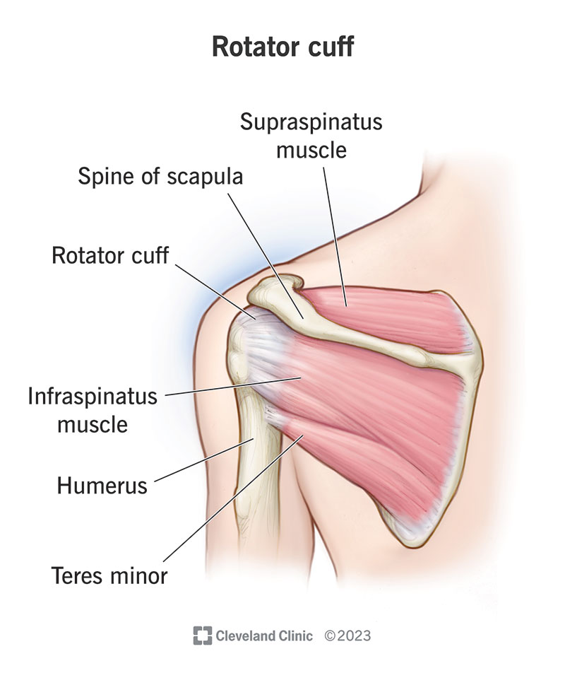
Rotator Cuff Muscles
The tendons of the rotator cuff muscles form a cuff around the shoulder joint on its anterior, superior, and posterior aspects. The muscles of the rotator cuff group are: - Supraspinatus - Infraspinatus - Teres minor - Subscapularis These muscles are all innervated by C5 and C6. Repeated abduction and flexion can cause wear and tear on the tendons, leading to cuff tears or rupture. The supraspinatus tendon is most vulnerable to injury.
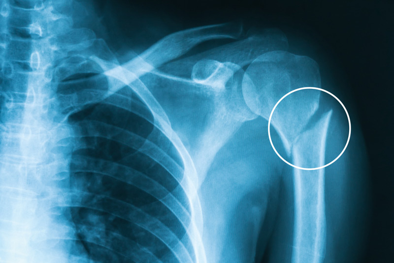
Humerus
Humeral Neck Fracture
These fractures often occur from a fall on an outstretched hand, especially common in elderly persons with osteoporosis. Key neurovascular structures at risk include the axillary nerve and posterior circumflex artery.
Shaft Fracture
Commonly associated with damage to the radial nerve due to its close relationship to the posterior aspect of the bone, running in the spiral groove.
Radial Nerve Palsy
This can result in 'wrist drop' from paralysis of the extensors of the wrist. There may also be sensory loss over the dorsal surface of the hand and the proximal ends of the lateral three and a half fingers dorsally.
Elbow
Joints and Injuries
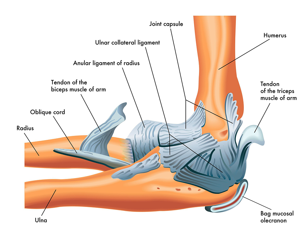
The elbow is a complex hinge joint allowing flexion, extension, and rotation of the forearm. The most common injuries include supracondylar fractures (especially in children) and radial head fractures (common in adults). Elbow dislocations often result from a fall on an outstretched hand.
Supracondylar Humeral Fractures
Injuries can cause damage to the anterior interosseous nerve (branch of the median nerve) and the brachial artery, leading to Volkmann's ischemic contracture of the forearm.
Forearm
Bones and Muscles
The forearm consists of the radius and ulna bones. Its muscles include the anterior compartment (flexors and pronators) and the posterior compartment (extensors).
Common Injuries
- Tennis Elbow: Overuse results in degeneration of the tendinous origin resulting in pain and inflammation at the lateral epicondyle.
- Golfer's Elbow: Similar to tennis elbow but affects the medial epicondyle.
- Monteggia and Galeazzi Fractures: Monteggia involves ulnar fracture and radial head dislocation, while Galeazzi involves radial fracture and distal radioulnar joint dislocation.
Wrist and Hand
Anatomy
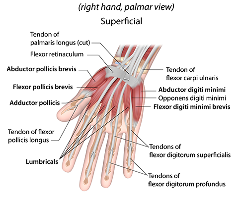
The wrist consists of multiple joints and bones, including the scaphoid, lunate, and others. Hand anatomy includes metacarpals and phalanges.
Common Injuries
- Distal Radial/Colles Fracture: Common in elderly osteoporotic women and caused by falling onto an outstretched hand.
- Scaphoid Fracture: May lead to avascular necrosis and non-union.
- Lunate Dislocation: Occurs from trauma to the wrist.
- Boxer's Fracture: Fracture of the metacarpal bones.
Nerves and Vessels
Brachial Plexus
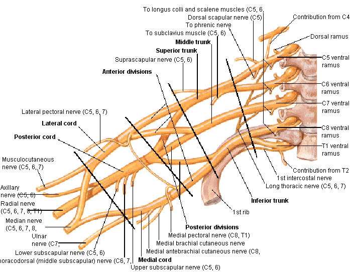
The nerve supply to the upper extremity arises from the brachial plexus (anterior rami of spinal nerves C5-T1). It is divided into roots, trunks, divisions, cords, and terminal branches. Injuries can cause conditions like Erb's Palsy (due to damage to the superior trunk) or Klumpke's Palsy (due to damage to the inferior trunk).
Radial Nerve
Injury to the radial nerve can occur at various levels, resulting in different deficits such as wrist drop.
Median Nerve
Injury at the elbow can cause difficulties in making a fist (hand of benediction). Carpal tunnel syndrome results from median nerve compression at the wrist.
ulnar nerve anatomy and injury impactsUlnar Nerve
Injuries can cause clawing of digits and impairments like Froment's sign.
Arteries and Veins
The arterial supply includes the subclavian, axillary, brachial, radial, and ulnar arteries. The venous return includes superficial and deep venous systems.