Abdominal Vasculature and Viscera
Anatomy of Abdominal Vasculature and Viscera
Intraperitoneal vs. Retroperitoneal Structures
*Note from an M2: You must memorize these two lists, unfortunately.
Intraperitoneal Organs:
- Stomach
- Liver
- Spleen
- Part of the duodenum
- Jejunum
- Ileum
- Transverse and sigmoid colon
Retroperitoneal Structures:
- Suprarenal glands
- Aorta and IVC
- Duodenum (2nd-4th parts)
- Pancreas (except tail)
- Ureters
- Ascending and descending colon
- Kidneys
- Esophagus
- Rectum (partial)
Esophagus
*Note from an M2: This isn't high yield. I would just know roughly where the aorta is, where the arteries are in relation to each other, which arteries are impacted if an organ anterior to it is inflamed etc...
- Constrictions:
- Cervical constriction
- Thoracic (aortobronchial) constriction
- Diaphragmatic constriction
- Anatomical Features:
- The transition from skeletal to smooth muscle in the middle third
- Gastroesophageal junction transition of mucosal lining
- Anterior and posterior vagal trunks accompany the esophagus

Stomach
Anatomical Features
- Regions:
- Cardiac
- Fundic
- Body
- Pyloric
- Curvatures:
- Greater curvature
- Lesser curvature
- Notable Structures:
- Cardiac notch
- Angular incisure
- Pyloric sphincter
- Gastric folds (rugae)
- Muscle Layers:
- Inner circular layer
- Outer longitudinal layer

Small Intestine
Sections
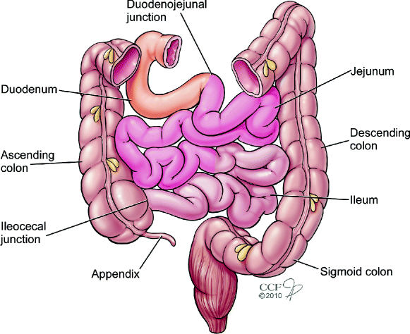
Duodenum:
- Duodenojejunal junction
- First few centimeters are smooth muscle
- Major and minor duodenal papillae

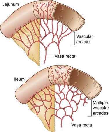
Jejunum:
- Occupies the upper left quadrant
- Less fat in mesentery
- Long, straight arteries and veins
- Abundant circular folds

Ileum:
- Occupies the lower right quadrant
- More fat in mesentery
- Shorter, straight arteries and veins
- Fewer circular folds
Functional Aspects
- Absorption of nutrients and water
- Total length is approximately 6 to 7 meters (22 feet)
Large Intestine
Features
- Larger diameter compared to the small intestine
- Retroperitoneal and Intraperitoneal Parts:
- Sacculation (haustra)
- Omental appendices (fat accumulations)
- Three longitudinal muscle bands (tenia coli)
Specific Regions
- Ileocecal Region:
- Contains the ileocecal junction
- Vermiform appendix with its own arterial supply (appendicular artery)
*Note from an M2: Know the arterial supply of different parts of the colon. They love anastomosis questions.

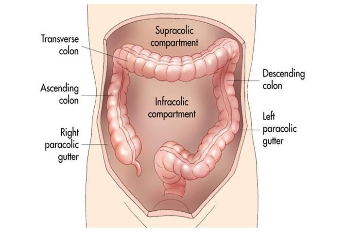
- Paracolic Gutters:
- Lateral to ascending and descending colon
- Blood-free mobilization possible by cutting peritoneum along these gutters

Blood Supply of Abdominal Viscera
Major Arteries
Celiac Trunk (Foregut):
- Supplies abdominal esophagus, stomach, duodenum (up to major duodenal papilla), liver, pancreas, gallbladder, and spleen
Superior Mesenteric Artery (Midgut):
- Supplies duodenum (part), jejunum, ileum, cecum, appendix, ascending colon, and transverse colon
Inferior Mesenteric Artery (Hindgut):
- Supplies descending colon, sigmoid colon, rectum, and upper part of the anal canal
Note from an M2: You will need to know the blood supply to different parts of the digestive system.

Anastomoses
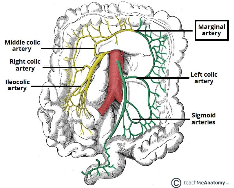
- Marginal Artery:
- Formed by anastomoses between right, middle, and left colic arteries
- Provides collateral circulation in case of inferior mesenteric artery stenosis

Nerve Supply of Abdominal Viscera
- Autonomic Nerve Fibers:
- Course with arteries
- Include both sympathetic and parasympathetic fibers
:background_color(FFFFFF):format(jpeg)/images/library/14062/gnEvUC2kgTFXvmrjP39Yw_Sympathetic_nervous_system.png)
Specific Plexuses
- Aortic Plexus
- Superior Hypogastric Plexus
- Hypogastric Nerve
- Pelvic Splanchnic Nerves
- Inferior Hypogastric Plexus

Clinical Considerations
- Duodenal Ulcers:
- Often occur in the superior part (duodenal cap)
- Posterior ulcers may erode onto major arteries, causing severe bleeding

