Anterior and Medial Thigh
Anterior and Medial Thigh
The lower limb plays pivotal roles in supporting body weight, enabling locomotion, and maintaining balance. The key structures involved in these functions include the femur, tibia, and a variety of muscles and nerves.
Lower Limb Functions
- Center of gravity
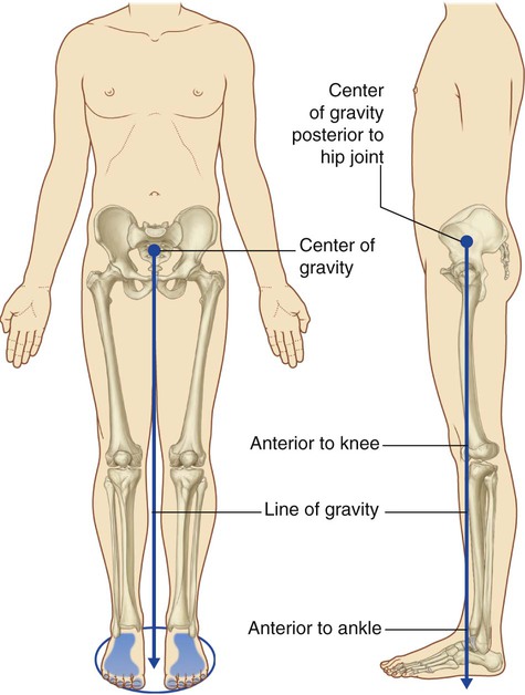
- Weight-bearing
- Locomotion
- Maintenance of balance
Bones and Regions of the Lower Limb
- Iliac crest: Top edge of the pelvis.

- Anterior superior iliac spine: Bony projection on the iliac crest.
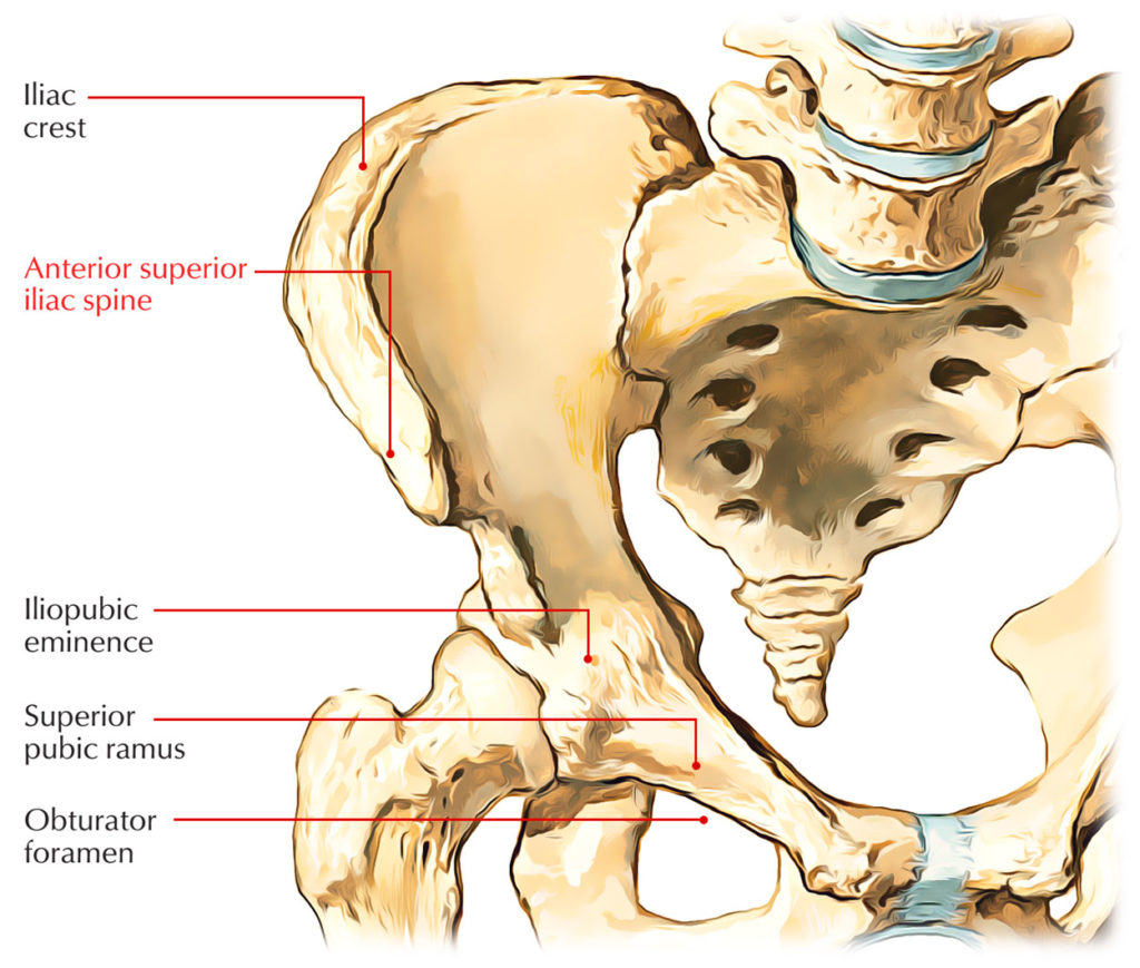
- Pubic symphysis: Cartilaginous joint uniting the left and right pubic bones.

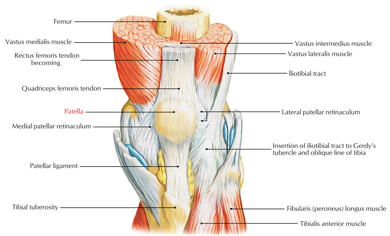
- Patella: Kneecap, a sesamoid bone.
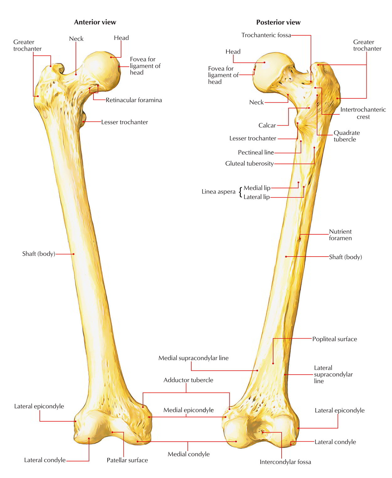
- Femur: The longest bone in the body.
- Tibia: The shin bone.
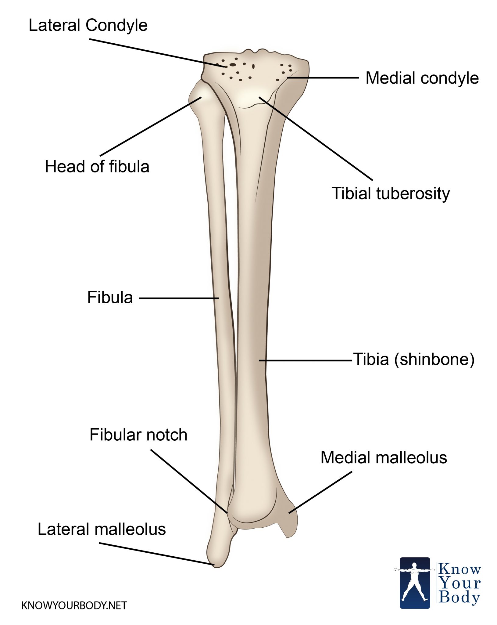
There are six major regions in the lower limb:
- Gluteal region

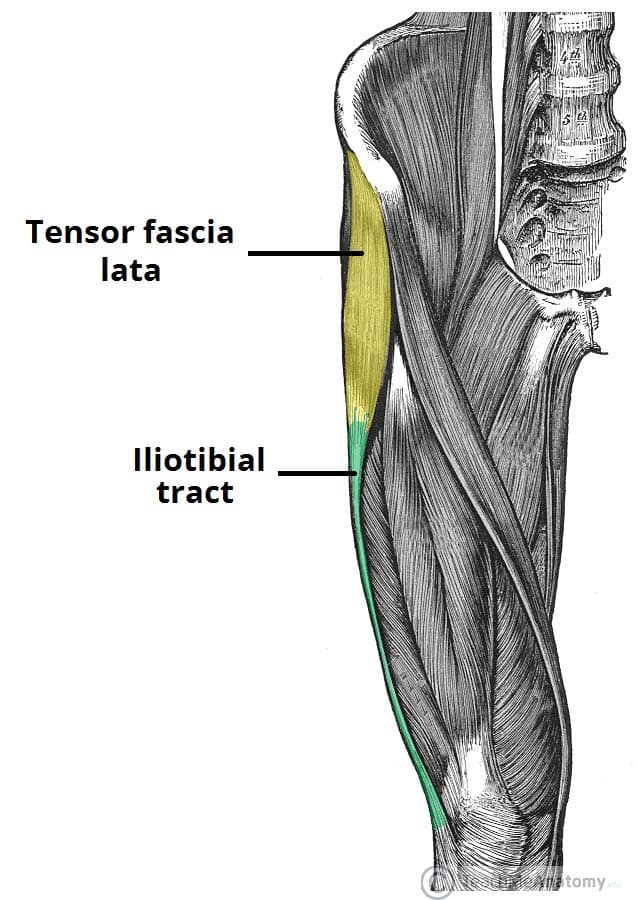
- Femoral region
- Knee region
- Leg region
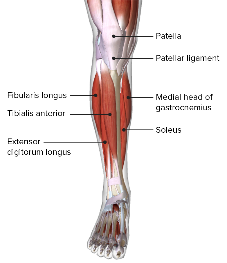
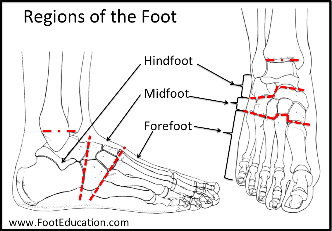
- Ankle region

- Foot region
Cross-Section of the Thigh
- Lateral Femoral Cutaneous Nerve

- Deep Fascia (Fascia Lata)
- Superficial and Deep Veins: The great saphenous vein is particularly notable.

Superficial Veins of the Lower Limb
- Great Saphenous Vein: The longest superficial vein.
- Small Saphenous Vein: Terminates at the popliteal vein.
Cutaneous Nerves of the Lower Limb
- Lateral Cutaneous branch of subcostal nerve
- Ilioinguinal Nerve
- Femoral branch of genitofemoral nerve
- Lateral Femoral Cutaneous Nerve
- Posterior Femoral Cutaneous Nerve
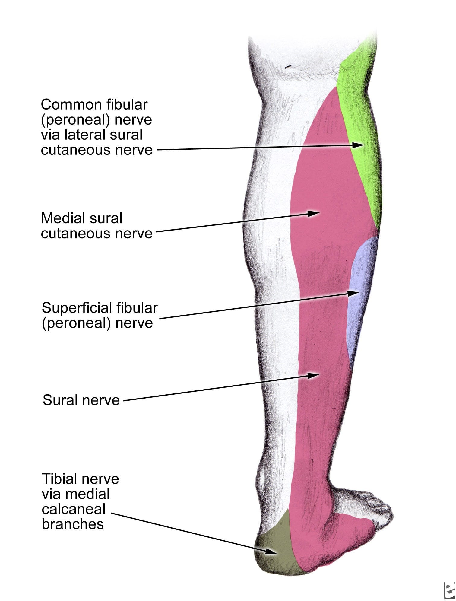
- Saphenous Nerve
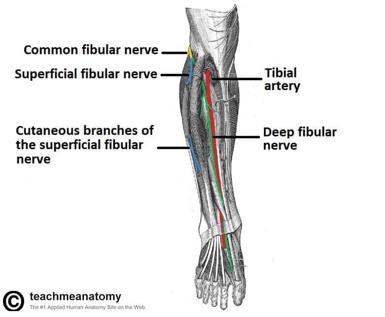
- Superficial and Deep Fibular Nerves
- Sural Nerve
Dermatomes of the Lower Limb
Anterior View and Posterior View: Represents the areas of the skin supplied by specific spinal nerves. 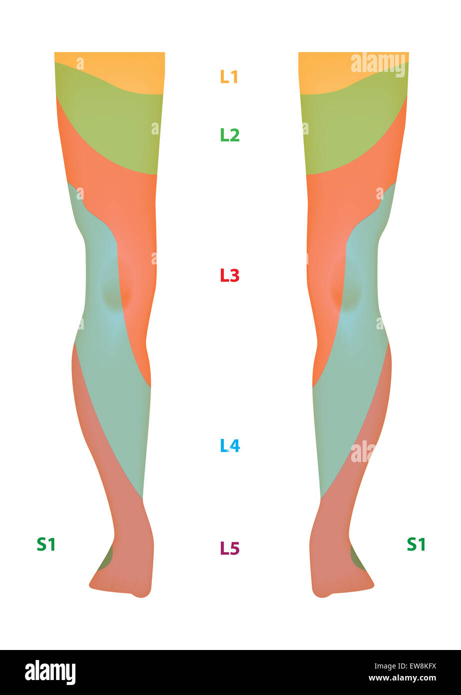
Lymphatics of the Lower Limb
- External Iliac Lymph Nodes
- Deep and Superficial Inguinal Lymph Nodes: The superficial group drains into the femoral canal.
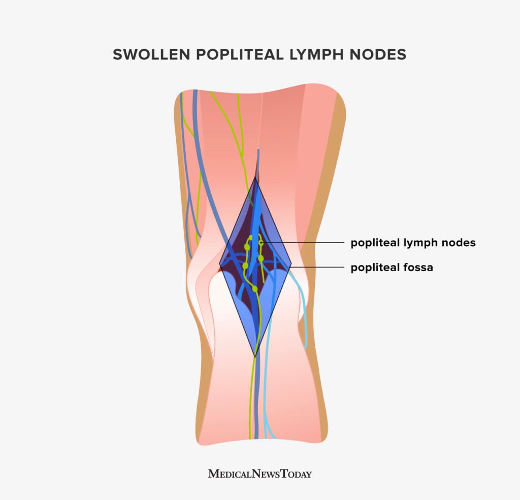
- Popliteal Lymph Nodes: Drain into the deep inguinal lymph nodes.
Femur
- Anterior View: Greater and lesser trochanter, shaft, lateral and medial condyle.
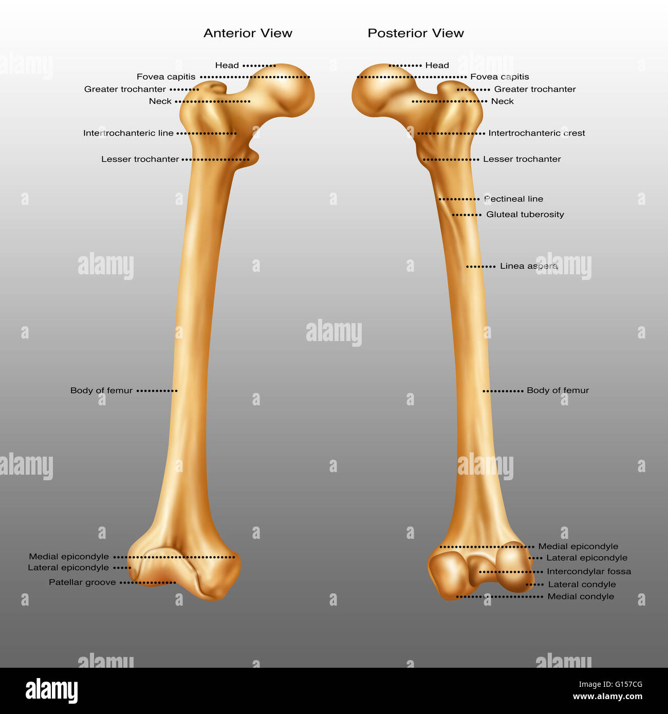
- Posterior View: Linea aspera, medial supracondylar line, adductor tubercle.
Femoral Triangle
Boundaries: Inguinal ligament, sartorius, adductor longus.
Contents: Femoral nerve, artery, and vein (NAVL configuration). 
Superficial Muscles of the Thigh
- Anterior Compartment: Iliopsoas, pectineus, sartorius, quadriceps femoris.
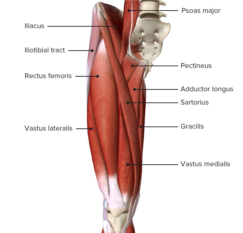
- Medial Compartment: Gracilis.
Deeper Muscles and Nerves of the Thigh
- Vastus Medialis, Intermedius, Lateralis, Rectus Femoris: Part of the quadriceps femoris.

- Femoral Nerve, Artery & Vein: Provide innervation and blood supply to the anterior compartment.

Arteries of Thigh and Knee
- Femoral Artery: Branches into the deep femoral artery.
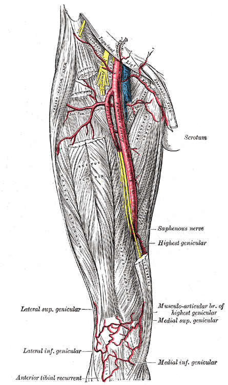
- Deep Femoral Artery: Perforating branches supply blood to various compartments.
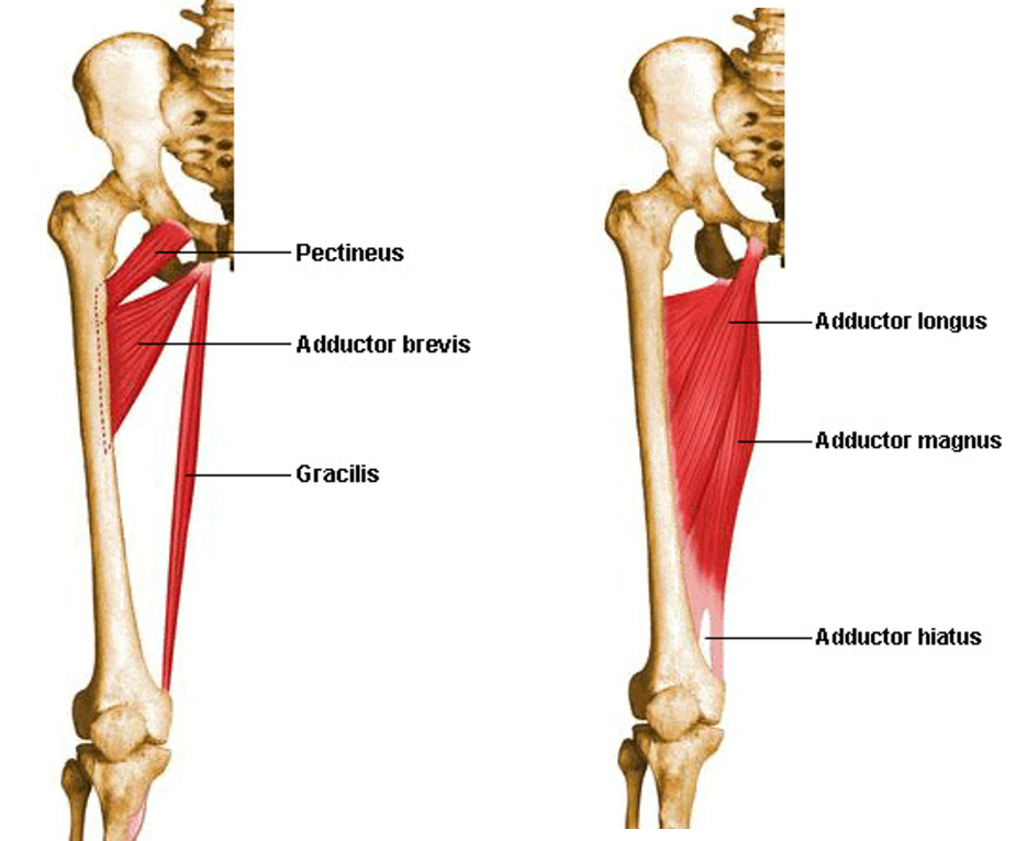
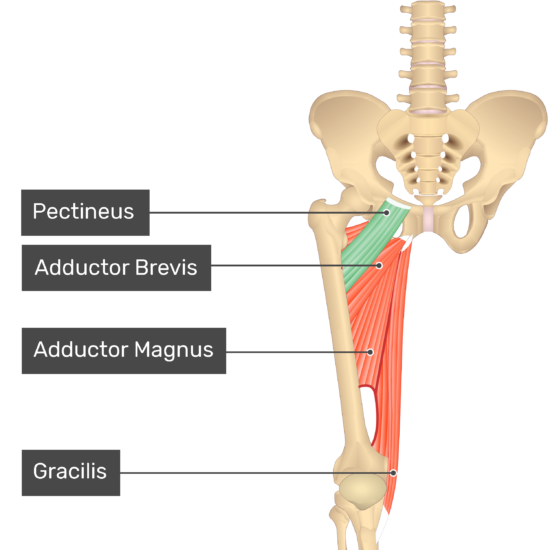
Muscles of the Medial Compartment of the Thigh (Adductor Group)
- Adductor Longus, Brevis, Magnus: Key adductor muscles.

- Pectineus and Gracilis: Assist in adduction and flexion at the hip joint.
Nerves and Arteries of the Medial Compartment
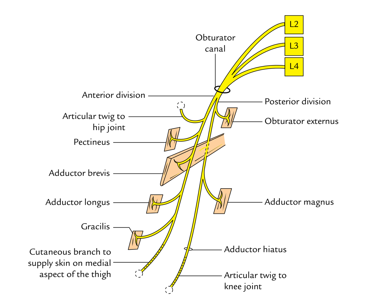
- Obturator Nerve: Innervates the adductor muscles.
- Deep Femoral and Obturator Arteries: Major blood suppliers.

These details provide a comprehensive overview of the anatomical structures and their functions within the anterior and medial regions of the thigh.