Shoulder Anatomy and Physiology
Shoulder Anatomy and Physiology
Bones of the Shoulder Region
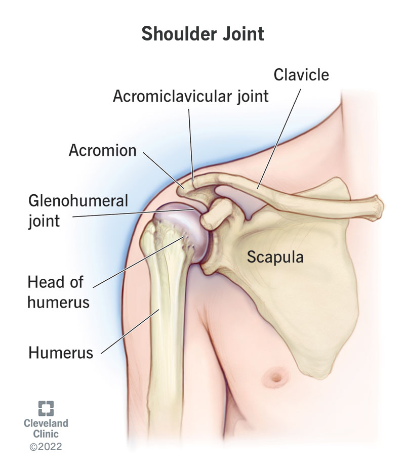
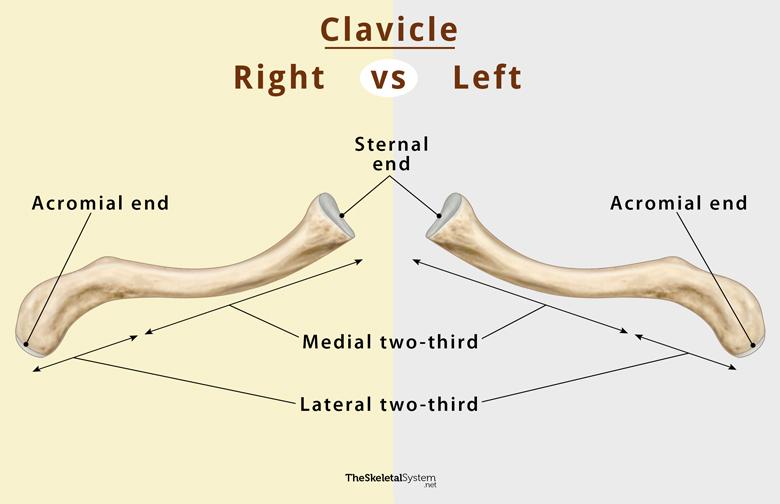
- Clavicle: Provides osseous continuity between the upper limb and the thorax, serves as a strut, and transmits traumatic impacts from the upper limb to the axial skeleton. It is palpable throughout its entire length and is one of the most commonly fractured bones.
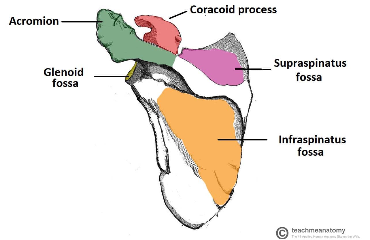
- Scapula: Key features include:
- Angles: Lateral, superior, and inferior.
- Borders: Superior, lateral, and medial.
- Processes: Acromion, spine, and coracoid.
- Surfaces: Costal and posterior.
Scapulohumeral Muscles
- Deltoid:
- Function: Major muscle responsible for the abduction of the arm.
- Nerve Supply: Axillary nerve.
- Blood Supply: Posterior circumflex humeral artery.
Supraspinatus
Rotator Cuff
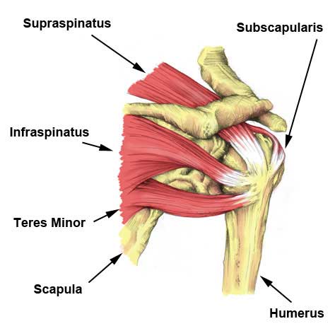
The rotator cuff consists of four muscles that stabilize the glenohumeral joint and allow for a wide range of shoulder movements:
- Supraspinatus: Most commonly involved in impingement and tendinopathy.
- Infraspinatus
- Teres Minor
- Subscapularis
:max_bytes(150000):strip_icc()/the-rotator-cuff-2696385-FINAL1-474e476cc4554dbd97995610f4402577.png)
Joint Structures and Ligaments
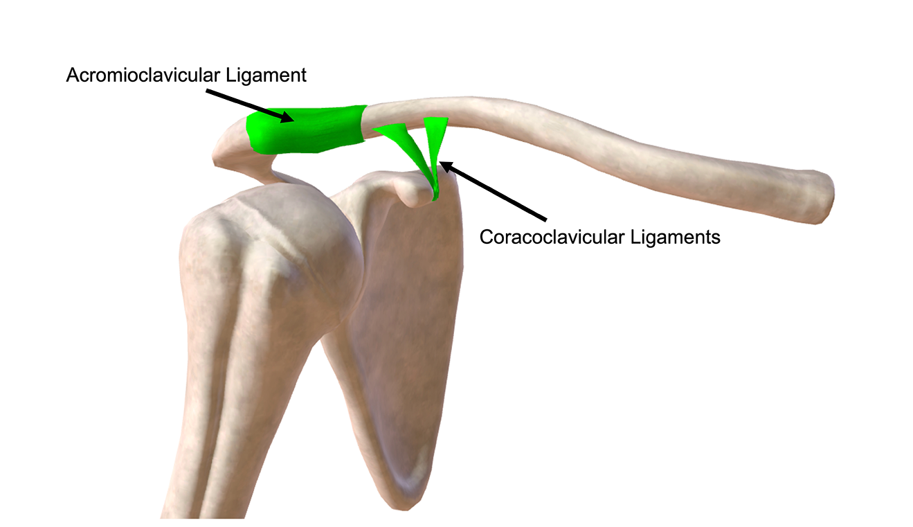
- Acromioclavicular Joint:
- Ligaments: Trapezoid, conoid, coracoclavicular, and acromioclavicular ligaments.
- Dislocation: This can occur due to trauma, with minor injuries tearing the fibrous joint capsule and more severe trauma disrupting the coracoclavicular ligament, leading to acromioclavicular separation.
- Glenohumeral Joint:
- Characteristics: A ball-and-socket synovial joint with a wide range of movements.
- Ligaments: Capsular ligament, coracohumeral ligament, transverse humeral ligament.
- Tendons: Subscapularis, supraspinatus, biceps (long head).
- Factors Stabilizing: Glenoid labrum, rotator cuff, coracoacromial arch, long head of biceps, long head of triceps.
- Common Dislocation: Most commonly dislocates in the inferior direction, clinically described as anterior or anteroinferior.
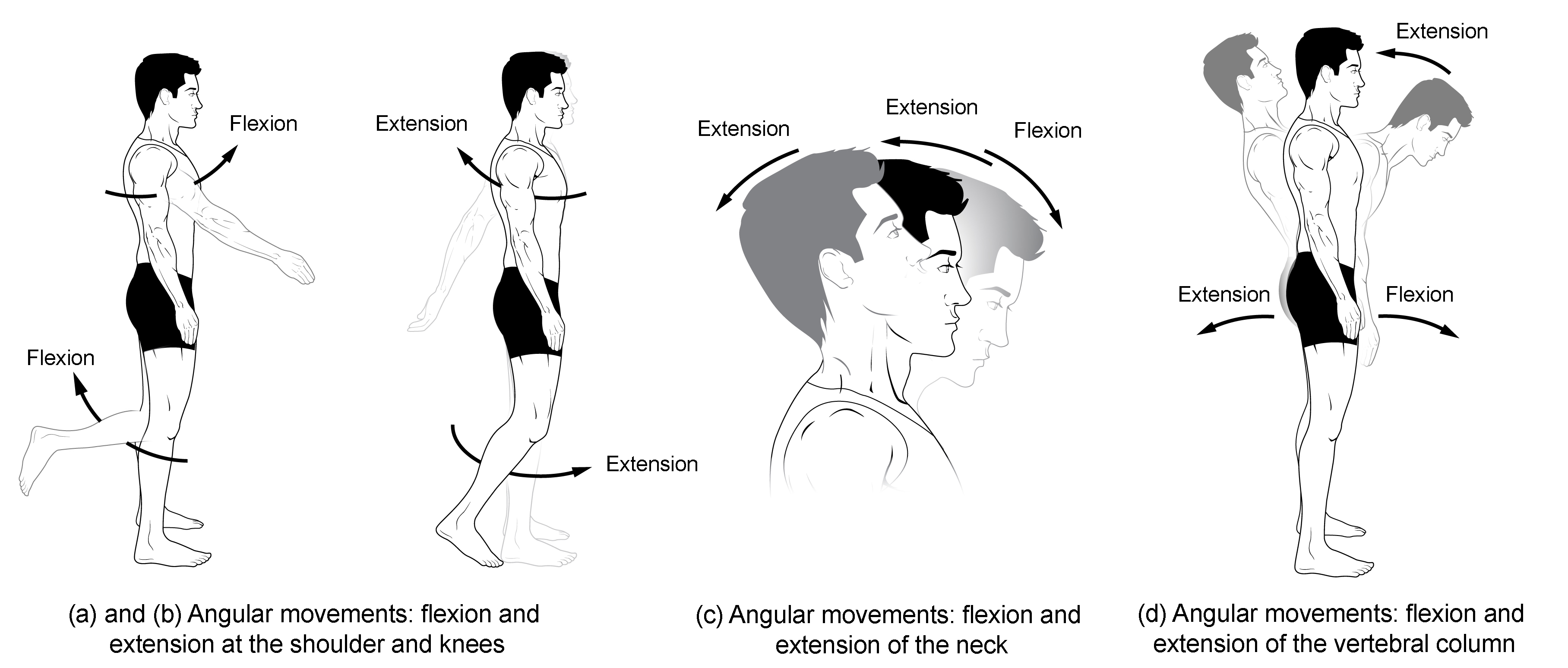
Movements of the Glenohumeral Joint
- Flexion and Extension: Movement along the medial-lateral axis.
- Abduction and Adduction: Movement along the antero-posterior axis.
- Medial and Lateral Rotation: Rotation around the vertical axis.
- Circumduction: A combination of all these movements allowing a full range of motion in multiple directions.
Arterial Anastomoses Around the Shoulder
The shoulder region is richly supplied with arterial anastomoses, ensuring adequate blood supply to the muscles and joints even in cases of partial vessel obstruction.
This detailed understanding of the shoulder region's anatomy and physiology provides the foundation for comprehending its function, joint injuries, and clinical relevance.
Here's a fun song to help you remember the content:
