Imaging Modalities and Anatomy of the Head and Neck
Imaging Modalities and Anatomy of the Head and Neck
Head & Neck Anatomy
- Plain Film (X-ray):
Role: Basic 2D imaging of bones, chest, and abdomen
Importance: Fast, low-cost, widely available; often the first-line imaging test - Ultrasound:
Role: Real-time imaging of soft tissues and blood flow
Importance: Non-ionizing, portable, cost-effective; excellent for obstetrics and vascular studies - CT (Computed Tomography):
Role: Detailed 3D imaging of entire body
Importance: Rapid, high-resolution; crucial for trauma, cancer staging, and complex diagnoses - MR (Magnetic Resonance):
Role: High-contrast soft tissue imaging without radiation
Importance: Superior for neurological, musculoskeletal, and certain organ studies; allows functional imaging - Fluoroscopy:
Role: Real-time X-ray imaging of moving body parts
Importance: Essential for guiding procedures and studying dynamic processes (e.g., swallowing studies) - Catheter Angiography:
Role: Detailed imaging of blood vessels
Importance: Gold standard for vascular imaging; allows for simultaneous diagnosis and treatment - Image-guided Procedures:
Role: Using various imaging modalities to guide minimally invasive interventions
Importance: Enables precise, less invasive treatments; reduces complications and recovery time




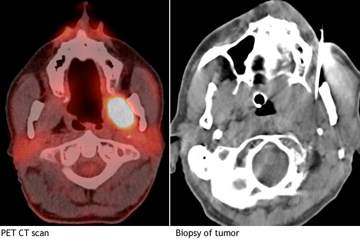
Neck
Suprahyoid and Infrahyoid Neck Spaces
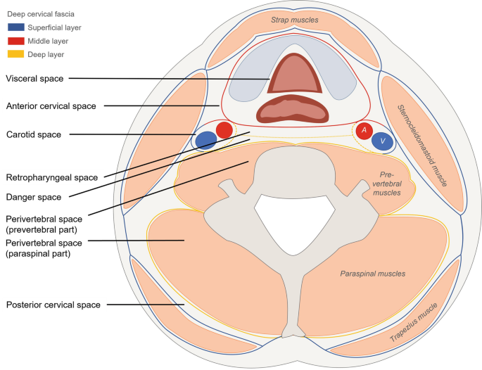
The neck spaces are divided by three layers of deep cervical fascia: - Superficial (investing) layer - Middle layer - Deep layer
Superficial Cervical Fascia
The superficial cervical fascia is a thin layer of subcutaneous connective tissue between the skin's dermis and deep fascia.
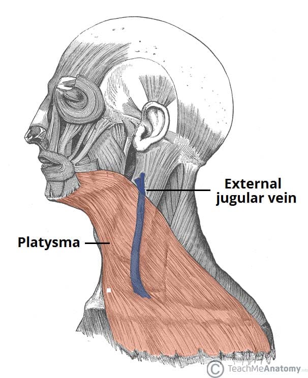
Deep Cervical Fascia
- Superficial layer: Investing fascia/masticator fascia/submandibular fascia/sternocleidomastoideoplazius fascia
- Middle layer: Strap muscle fascia and visceral fascia
- Deep layer: Perivertebral fascia (prevertebral for the anterior part only)
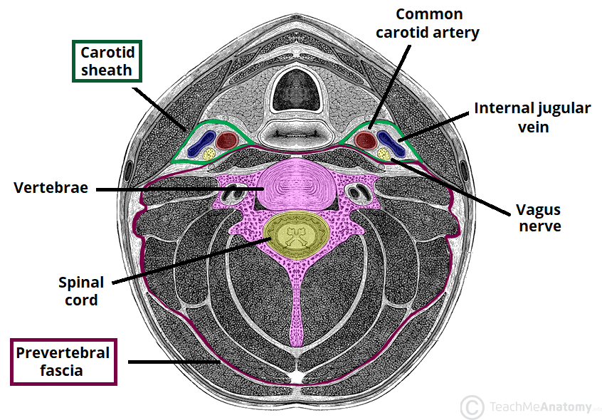
Imaging Techniques
X-Ray of the Neck
An X-ray of the neck can reveal important anatomical landmarks, such as:
-
Middle Turbinate
- Inferior Turbinate
- Adenoid
- Cervical Body 4
- Intervertebral Disk C4
- Soft Palate
- Superior Articular Process C5
- Tongue
- Genioglosses Muscle
- Tonsil
- C6 Trachea
- Spinous Process C7
- Inferior Articular Process
- Mandible
- Thoracic Body 1
- Hyoid Bone
- Epiglottis
- Thyroid Cartilage
-Vocal Cords
- Trachea
- Esophagus

CT of the Neck
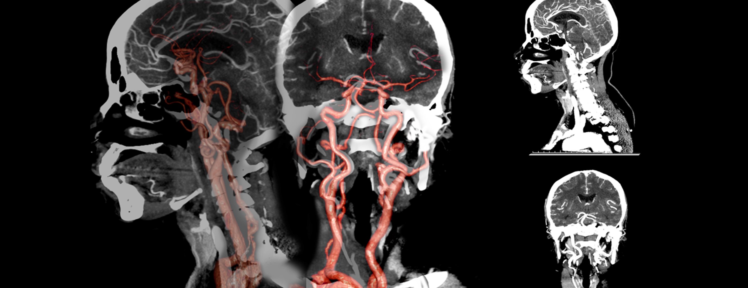
CT is the imaging modality of choice for neck imaging and is performed with IV iodinated contrast. It covers from the frontal sinus to the aortic arch, utilizing a single breath hold technique and multiplanar reconstruction with different window settings (soft tissue and bone).

Indications for CT Neck: - Inflammatory or infectious processes - Head and neck neoplasms - Head and neck cancer therapy response and follow up - Head and neck trauma - Vascular malformations - Bleeding/epistaxis - Congenital anomalies - Foreign bodies - Thyroid disease
Anatomy Highlighted in CT Scans
- Globe
- Optic Nerve
- Eustachian Tube
- Posterior Communicating Artery
- Levator Veli Palatini Muscle
- Parapharyngeal Fat
- Masseter Muscle
- Longus Coli Muscle
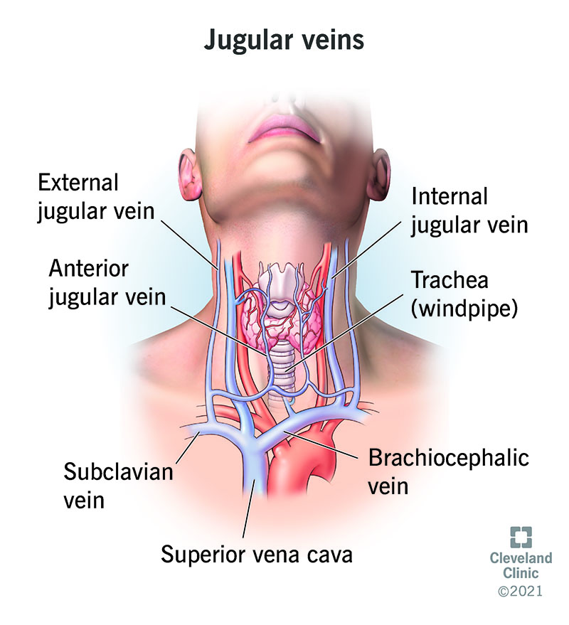
- Jugular Vein
- Occipital Artery
- Mylohyoid Muscle
- Oropharynx
- Lingual Artery
- Vallecula
- Epiglottis
- Submandibular Gland
- Trachea
- Esophagus
- Aortic Arch

Neck Spaces Overview
Suprahyoid Neck Spaces
Parapharyngeal Space: Contains fat, minor salivary glands, internal maxillary artery, ascending pharyngeal artery, pterygoid venous plexus, buccinator muscle, palatine tonsil, buccal space, mandibular ramus, masseter muscle, medial pterygoid muscle, retromandibular vein, deep lobe of the parotid gland, styloid process, carotid space, internal jugular vein, and internal carotid artery.
Pharyngeal Mucosal Space: Includes the mucosal surface of the pharynx, lymphatic ring, minor salivary glands, pharyngobasilar fascia, pharyngeal mucosal space muscles, and the torus tubarius.
Masticator Space: Contains muscles of mastication, mandibular nerve (V3), ramus & posterior body of the mandible, maxillary artery, pterygoid venous plexus.
Parotid Space: Houses the parotid gland, parotid duct, extracranial facial nerve (CNVII), external carotid artery, retromandibular vein, and intraparotid lymph nodes.
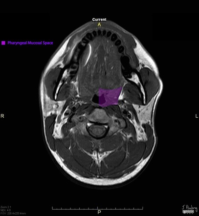


Infrahyoid Neck Spaces
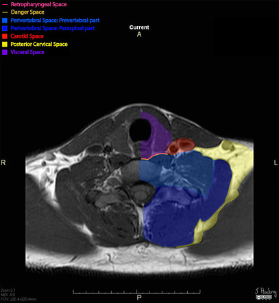
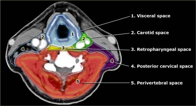
The infrahyoid neck is divided into several compartments:
Visceral Space: Contains structures such as the larynx, hypopharynx, trachea, esophagus, thyroid gland, parathyroid glands, lymph nodes, and recurrent laryngeal nerves.
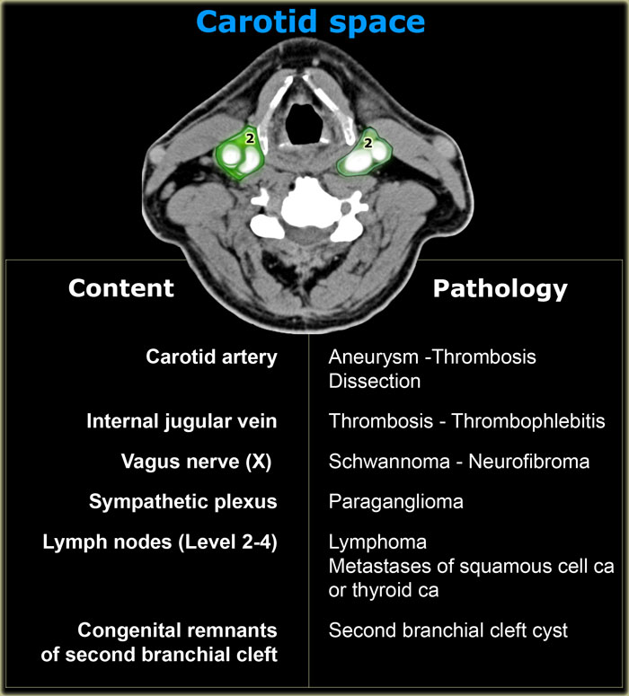
Carotid Space: Encompasses the common and internal carotid artery, internal jugular vein, vagus nerve (CNX), sympathetic trunk, and deep cervical chain lymph nodes.
Retropharyngeal Space: Potential space containing lymph nodes and fat, bordered by the prevertebral layer of deep cervical fascia.
Danger Space: A potential space that extends from the skull base to the mediastinum, posing the risk of deep infections spreading.
Key Neck Pathologies
- Abdominal Aneurysm: Thrombosis and dissection of the carotid artery.
- Jugular Vein Thrombosis: Thrombophlebitis and related complications.
- Vagus Nerve Schwannomas and Neurofibromas: Tumors of the nerve.
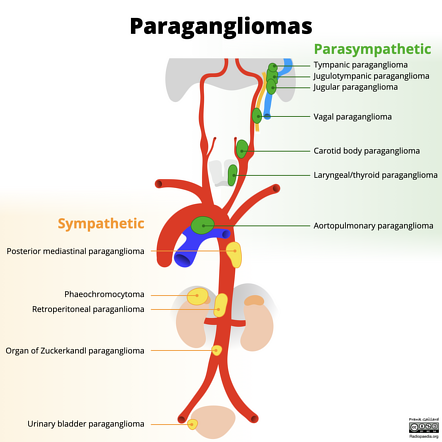
- Paraganglioma: Tumors involving the sympathetic plexus.
- Lymphatic Metastases: Seen in malignancies of the head and neck region.
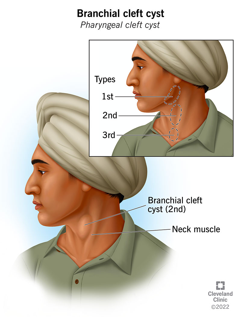
- Second Branchial Cleft Cyst: Congenital anomalies in the carotid space.

