Thyroid and Parathyroid Glands, Root of the Neck
Thyroid and Parathyroid Glands, Root of the Neck
Thyroid Gland
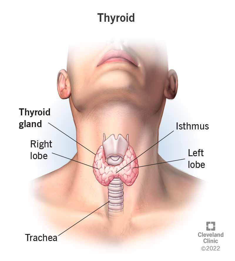
The thyroid gland is located in the neck and consists of two lobes - the right and left lobes - connected by an isthmus. The position of the thyroid gland is as follows:
- Hyoid bone
- Thyroid notch
- Laryngeal prominence
- Median cricothyroid ligament
- Arch of cricoid
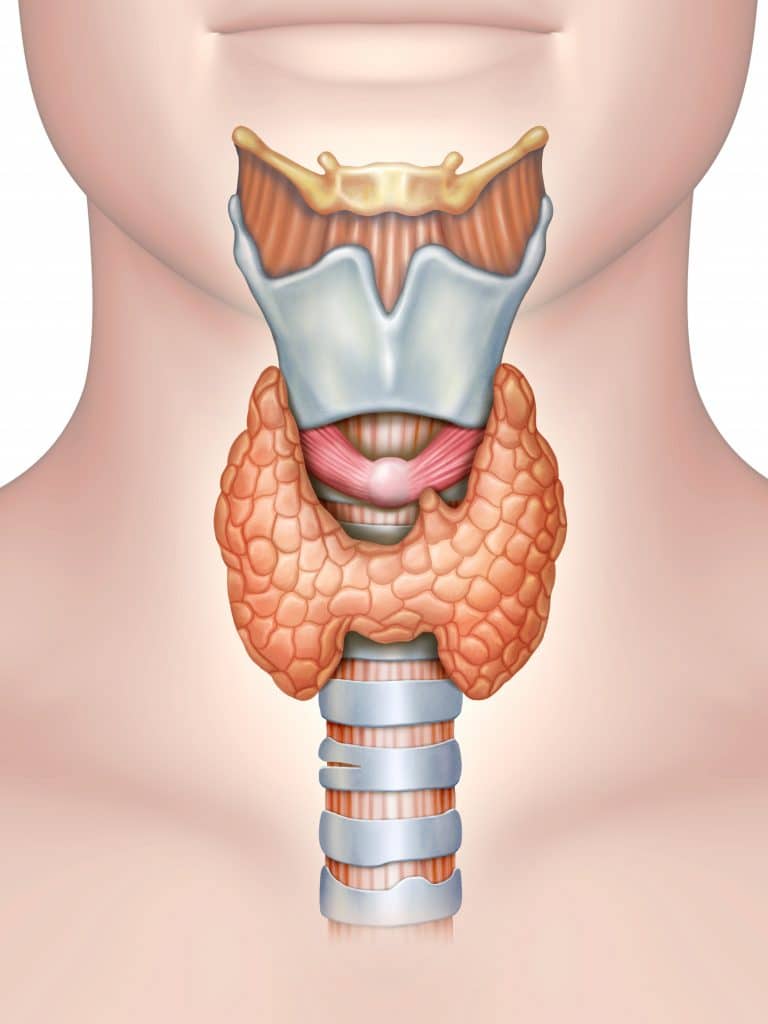
Structure
- Lobes: The thyroid gland has two lobes.
- Pyramidal lobe: Present in approximately 50% of individuals.
- Isthmus: Located over the 2nd to 4th tracheal rings.
Parathyroid Gland
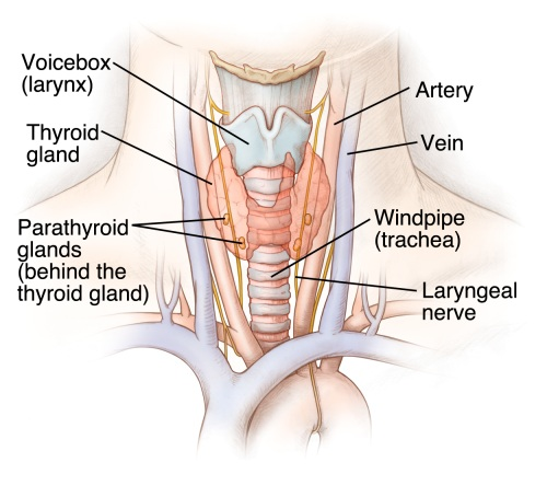
The parathyroid glands are small endocrine glands located on the posterior surface of the thyroid gland. There are typically two pairs (superior and inferior), with the following characteristics:
- Located between the capsule and the sheath of the thyroid gland.
- Each gland has its own capsule and vascular supply.
- The lower two glands can migrate during development, which may result in ectopic parathyroid glands.
Vasculature of the Thyroid Gland
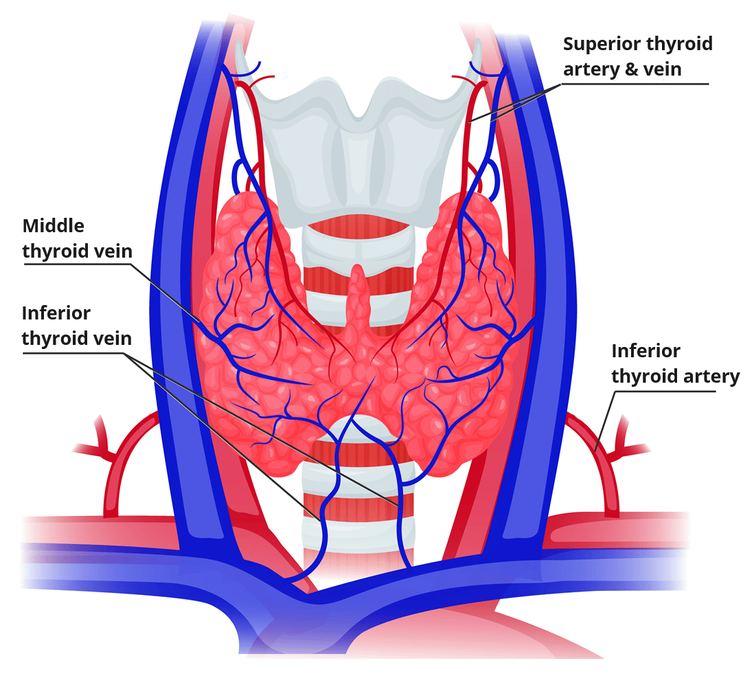
The thyroid gland is highly vascularized, with major arteries and veins supplying it:
- Superior thyroid artery: Branches into anterior and posterior glandular branches.
- Inferior thyroid artery: Supplies the inferior part of the gland.
- Thyroid ima (“lowest”) artery: Unpaired artery present in about 10% of individuals.
- Superior thyroid veins and middle thyroid veins: Drain into the internal jugular vein.
- Inferior thyroid veins: Drain into the brachiocephalic veins.
Nerves
Key nerves to be aware of during thyroid surgery or procedures include:
- Recurrent laryngeal nerve: Susceptible to injury during thyroid surgery.
- Superior laryngeal nerve: Provides sensation to the larynx.
:background_color(FFFFFF):format(jpeg)/images/article/thyroid-gland/7Ax0xdjleIEG3N69hNgksA_IlX2fetaanDEiPz7vGWACw_Thyroid_1.png)
Root of the Neck
The root of the neck is a crucial area connecting the head, neck, thorax, and upper limb. Components include:
Contents
- Carotid sheath: Encloses the carotid artery, internal jugular vein (IJV), and vagus nerve (CNX).
Vessels
- Subclavian artery and vein: Key vessels traversing this area.
- Thoracic duct and lymphatics: Major lymphatic vessels present.
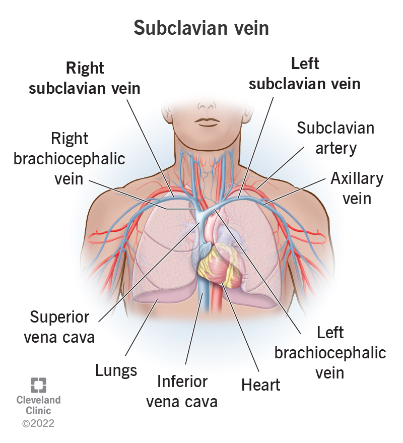
Nerves
- Phrenic nerve: Innervates the diaphragm.
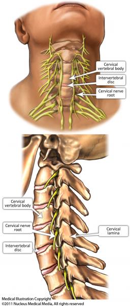
- Brachial plexus: Supplies the upper limb.
- Cervical sympathetic trunk: Sympathetic nerve fibers running through the neck.
Subclavian Artery
The subclavian artery is divided into three sections, defined relative to the anterior scalene muscle:
First Section
- Vertebral artery: Ascends through the transverse foramina of cervical vertebrae.
- Thyrocervical trunk: Has branches including the inferior thyroid artery.
- Internal thoracic artery: Descends into the thorax.

Second Section
- Costocervical trunk: Branches into the supreme intercostal artery and deep cervical artery.
Third Section
- Dorsal scapular artery: Supplies the scapular region, variable in origin.

Vertebral Artery
The vertebral artery passes through the transverse foramina of cervical vertebrae and enters the cranial cavity through the foramen magnum. It supplies the posterior part of the brain.
:background_color(FFFFFF):format(jpeg)/images/article/vertebral-artery/Pr3DUiOydFe8hJ3WXcIg_qZw8tXTIX5JHJ79oMmfKA_Vertebral_artery.png)
Sympathetics of the Neck
Cervical Sympathetic Trunk
The cervical sympathetic trunk includes the following ganglia:
- Superior cervical ganglion
- Middle cervical ganglion
- Inferior (cervicothoracic or stellate) cervical ganglion

Injury
Injuries to the sympathetic trunk can result in Horner syndrome, characterized by:
- Constricted pupil (miosis)
- Drooping eyelid (ptosis)
- Lack of sweating on the affected side (anhidrosis).
- Flushed complexion (vasodilation)
*Note from an M2: This will come up a lot in brain and behavior.
:max_bytes(150000):strip_icc()/horner-syndrome-symptoms-causes-diagnosis-and-treatment-4176967_FINAL-5c05c619c9e77c0001e609e8.png)
Lymphatics of the Root of the Neck
The lymphatic system in the root of the neck is vital for draining lymph from various body regions:
- Right lymphatic duct: Drains lymph from the right side of the thorax, right upper extremity, and right side of the head and neck.
- Thoracic duct: Drains lymph from the rest of the body, entering the root of the neck posterior to the carotid sheath.
Summary
The thyroid and parathyroid glands, along with key anatomical structures in the root of the neck, have critical functions and implications for surgical procedures and clinical conditions.