Thoracic Wall and Lungs
Thoracic Wall and Lungs
Notes from an M2:
Don't forget to know the boundaries of the thoracic cavity. I missed several points in this unit because I didn't memorize them. Know which structures are in which cavity, and know the boundaries on all sides. You need to understand how these organs are laid out in 3d. You may be asked which structure is anterior, posterior, or lateral and do a different structure. The first and second ribs are tested often. Know which structures you will encounter to start at the epidermis and go toward the inner organs. Know where foreign objects are more likely to lodge in bronchi.
Thorax Overview
Thoracic Boundaries & Skeleton: The thorax, also known as the chest, is an area of the body located between the neck and the diaphragm. Thanks to the thoracic skeleton, which is composed of the sternum, ribs, and thoracic vertebrae, it functions as a protective cage.
Superior & Inferior Thoracic Apertures:
- Superior Thoracic Aperture (Thoracic Inlet): The inlet allows communication between the thorax, neck, and upper limbs. It is bounded by the first thoracic vertebra posteriorly, the first pair of ribs laterally, and the superior border of the manubrium anteriorly.
- Inferior Thoracic Aperture (Thoracic Outlet): The outlet is closed by the diaphragm, separating the thoracic cavity from the abdominal cavity. It is bounded by the twelfth thoracic vertebra, twelfth pair of ribs, the costal margin, and the xiphisternal joint.

Thoracic Wall & Intercostal Muscles
Bones, Muscles, Nerves, Vessels, Parietal Lymphatics: The thoracic wall primarily comprises the thoracic vertebrae, ribs, and sternum. It serves as an attachment point for muscles and protects the thoracic organs. Key components of the thoracic wall include:
- Bones: Sternum (manubrium, body, xiphoid process), ribs (true ribs 1-7, false ribs 8-10, floating ribs 11-12).
- Muscles: External intercostal muscles, internal intercostal muscles, innermost intercostal muscles, subcostal muscles, transversus thoracis muscle.
- Nerves: Intercostal nerves originating from the thoracic spinal nerves.
- Vessels: Intercostal arteries and veins.
- Parietal Lymphatics: Lymph nodes and vessels responsible for draining the thoracic wall.
Endothoracic Fascia: The endothoracic fascia is a thin layer of loose connective tissue between the thoracic wall and the parietal pleura. It provides a cleavage plane, allowing surgeons to separate the parietal pleura from the thoracic wall to access intrathoracic structures.
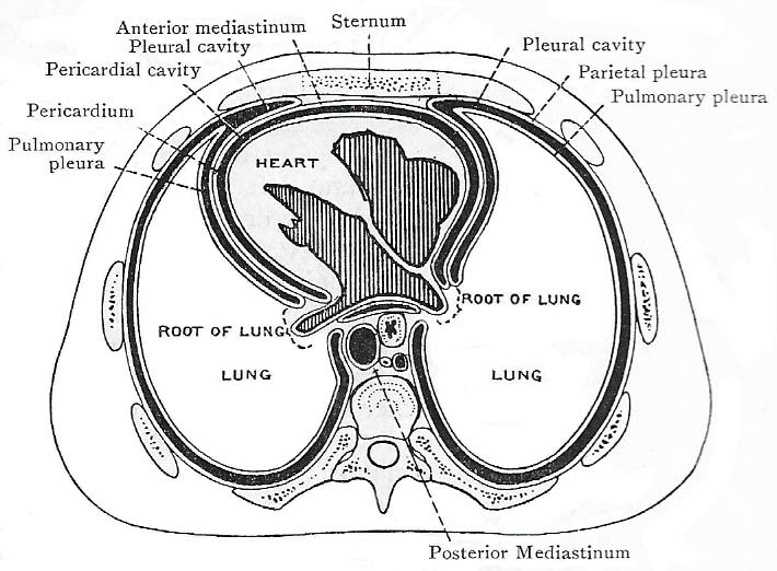
Thoracic Cavity
Subdivisions: The thoracic cavity is divided into three main components:
- Two Pleural Cavities: Each containing a lung, lined by pleurae (visceral and parietal).
- Mediastinum: The central compartment of the thoracic cavity, divided into superior and inferior mediastinum. It houses organs such as the heart, esophagus, trachea, great vessels, and nerves.

Pleura and Pleural Cavity
Pleura: The pleura is a serous membrane that envelops the lungs and lines the thoracic cavity. It consists of two layers:
- Visceral Pleura: Covers the lungs directly.
- Parietal Pleura: Lines the inner surface of the thoracic wall, mediastinum, and diaphragm. It is divided into cervical (cupula), costal, mediastinal, and diaphragmatic pleura.
Pleural Cavity: The pleural cavity is a potential space between the visceral and parietal layers that contains a small amount of serous fluid. This fluid reduces friction between the pleura during breathing movements.
Recesses and Reflections:
- Costodiaphragmatic Recess: A potential space at the junction of the costal and diaphragmatic pleura.
- Costomediastinal Recess: Located at the junction of the costal and mediastinal pleura.
- Pulmonary Ligament: A double-layered extension of the pleura that extends from the hilum of the lung to the diaphragm.

Lungs
The lungs are vital respiratory organs divided into lobes and segments, facilitating gas exchange.
- Right Lung: Divided into three lobes (superior, middle, and inferior) by the oblique and horizontal fissures.
- Left Lung: Divided into two lobes (superior and inferior) by the oblique fissure. The left lung also features the cardiac notch and the lingula.
Trachea and Bronchi
The trachea bifurcates at the level of the sternal angle into the right and left primary bronchi. Key characteristics include:
- Right Bronchus: Shorter, wider, more vertical. More prone to aspiration of foreign bodies.
- Left Bronchus: Longer and more angled compared to the right.

Lung/Mediastinal Relationships
The right lung is related to structures such as the superior vena cava, azygos vein, and esophagus, while the left lung is related to the heart, aortic arch, thoracic aorta, and esophagus.
Bronchopulmonary (BP) Segments
Definition: A BP segment is the largest subdivision of a lung lobe, pyramidal in shape, and supplied by a segmental (tertiary) bronchus and accompanying pulmonary artery branch.
Characteristics:
- Largest subdivision of a lobe.
- Separated by connective tissue septa.
- Surgically resectable.
- Drained by intersegmental parts of pulmonary veins.
:watermark(/images/watermark_only_413.png,0,0,0):watermark(/images/logo_url_sm.png,-10,-10,0):format(jpeg)/images/anatomy_term/superior-segment-of-left-lung/n9rHAj2ECgYaeXGP21j8nQ_Superior.png)
*Note: It's important to distinguish between the red and blue. The substructures themselves are low-yield.
Cadaveric Impressions and Structures in the Hila
Right Lung Cadaveric Impressions: Grooves for the superior vena cava, esophagus, and trachea are noted. The hilum contains structures such as the right pulmonary artery, pulmonary veins, and primary bronchus.
Left Lung Cadaveric Impressions: Grooves for the descending aorta and subclavian artery. The hilum structures include the left pulmonary artery, veins, and primary bronchus.

*Note: You're bound to get a couple questions about the hillum.
Phrenic Nerve
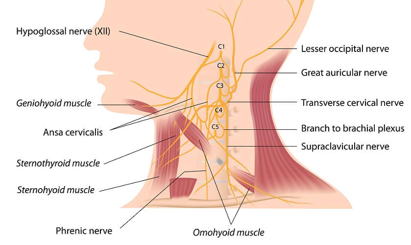
The phrenic nerve (C3, C4, and C5) innervates the diaphragm. It provides motor and sensory innervation to the diaphragm and parts of the pleura.

Hemothorax and Thoracentesis
Hemothorax: Accumulation of blood in the pleural cavity, potentially compressing the lung.
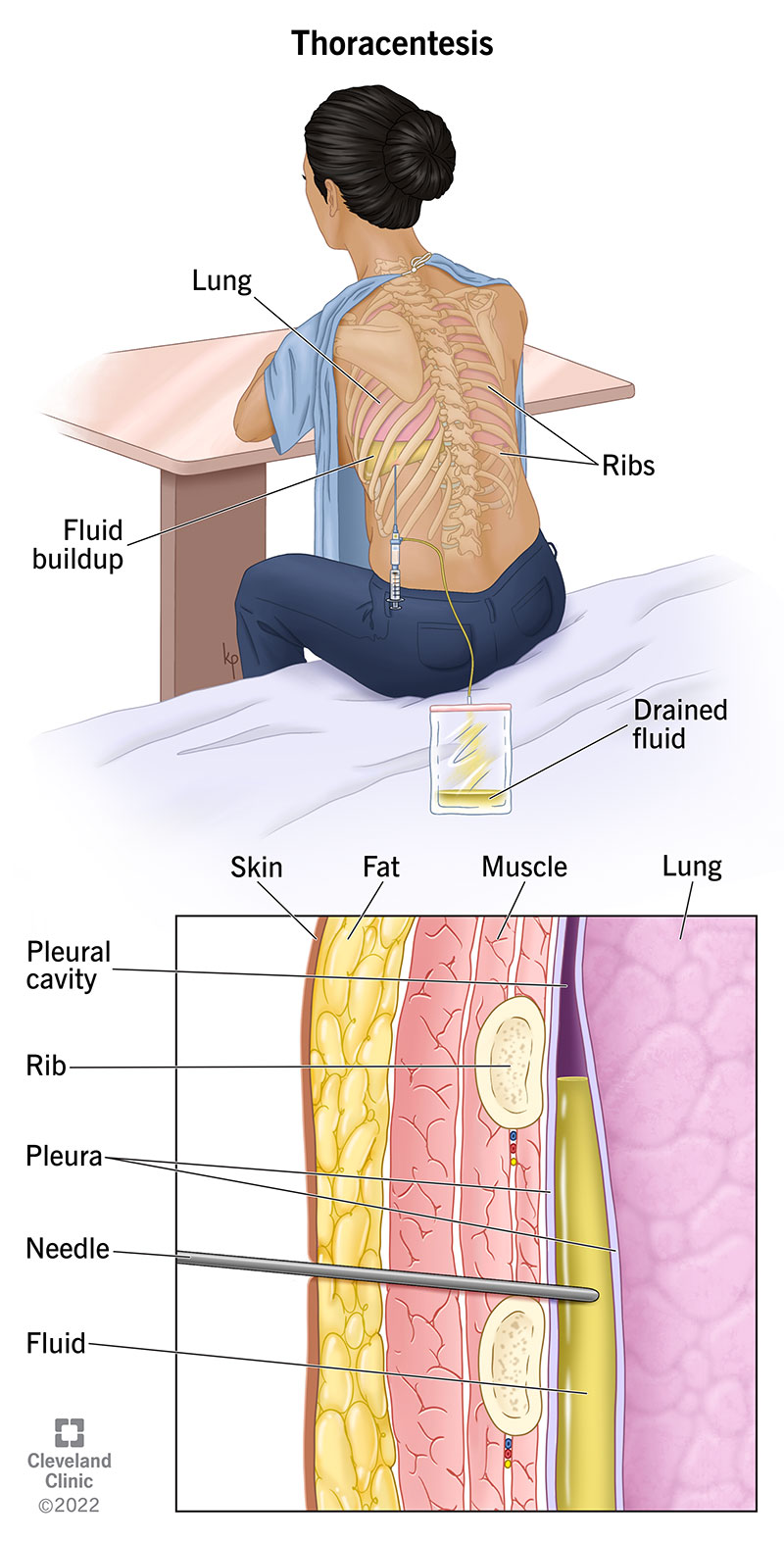
Thoracentesis: A procedure to remove fluid from the pleural cavity. It is typically performed in the midaxillary line, avoiding major vessels and nerves.
Dissection
Dissection involves removing the anterior thoracic wall to study internal structures. Care is taken to cut and reflect various muscles, arteries, and ribs for detailed examination.
Here's a helpful song:
