Temporal Region and Infratemporal Fossa
Temporal Region and Infratemporal Fossa
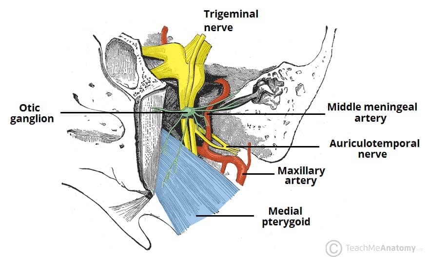
Infratemporal Fossa
- Contents:
- Inferior part of the Temporalis muscle
- Lateral and medial pterygoid muscles
- Maxillary artery
- Pterygoid venous plexus
- Mandibular, inferior alveolar, lingual, buccal, and chorda tympani nerves
- Otic ganglion
- Covered by the parotid gland externally
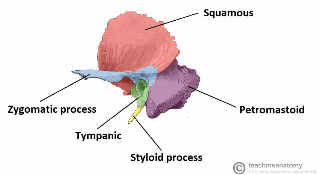
Osteology of the Temporal Bone
- Articular fossa
- Articular tubercle
- External acoustic meatus (opening for outer ears)
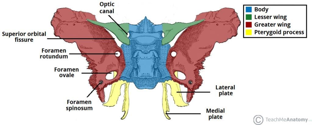
Sphenoid Bone
- Sphenoidal sinus
- Medial pterygoid plate
- Greater wing
- Sphenoid body
- Lateral pterygoid plate
Mandible
- Pterygoid fovea (neck)
- Condylar process (head) that sits in the auricular fossa
- Coronoid process which serves as insertion for the temporalis muscle
- Mandibular notch
- Mandibular foramen (entry point for inferior alveolar nerve/vessels to supply the lower mouth)
- Ramus
- Mental foramen
- Angle of the mandible

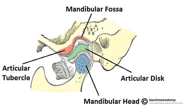
Temporomandibular Joint (TMJ)
- Articular tubercle
- Mandibular fossa
- Coronoid process
- Condylar process
Temporomandibular Joint (TMJ) Information:
- Synovial Joint
- Articular surfaces covered with fibrocartilage
- Articular Disc
- Pars Meniscus (attached to lateral pterygoid muscle)
- Pars Gracilis (poorly innervated and vascularized)
- Pars Posterior (thickest)
- Joint Capsule
- Fascial thickenings and extracapsular ligaments:
- Lateral ligament
- Stylomandibular ligament
- Sphenomandibular ligament
Movements of TMJ
- Mouth Closed
- Mouth opened past 15 degrees (lateral pterygoid translates the head of the mandible and articular disc anteriorly)
- Depression (gravity-assisted by suprahyoid and infrahyoid muscles to depress mandible)
- Protrusion (lateral pterygoid with assist from medial pterygoid)
- Elevation (closing the mouth assisted by temporalis, masseter, and medial pterygoid)
- Retrusion (posterior fibers of temporalis, deep part of masseter, geniohyoid, and anterior belly of digastric)
- Lateral movement (grinding and chewing utilizing temporalis, masseter, and pterygoids)
- Dysfunction and dislocations of TMJ joint disorders
Muscles of Mastication
Temporalis and Masseter Muscles
- Temporalis
- Temporal fascia and muscle
- Innervated by trigeminal nerve [V3]
- Masseter
- Masseteric nerve and artery
- Innervated by trigeminal nerve [V3]
Medial and Lateral Pterygoid Muscles
- Medial Pterygoid Muscle
- Mirrors the masseter; elevates, protrudes, moves mandible laterally/grinds
- Innervated by trigeminal nerve [V3]
- Lateral Pterygoid Muscle
- Opens jaw (depresses mandible)
- Both heads insert into the capsule of TMJ
- Innervated by trigeminal nerve [V3]
- Clinical note: Weakness in CN V3 causes jaw deviation to the affected side when opening/closing the mouth
:background_color(FFFFFF):format(jpeg)/images/library/14075/Masticatory_muscles.png)
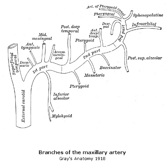
Maxillary Artery
- Branches
- Deep temporal arteries
- Middle meningeal artery
- Buccal artery
- Inferior alveolar artery
Pterygoid Plexus
- Connections
- Emissary veins
- Inferior ophthalmic vein (connect with cavernous sinus)
- Superficial temporal vein
- Maxillary vein
- Deep facial vein
- Retromandibular vein
- Inferior alveolar vein
- Posterior auricular vein
- Facial vein
- External jugular vein
- Internal jugular vein

Trigeminal Nerve (CN V3 - Mandibular Branch)
Motor Nerves
- Nerve to mylohyoid, anterior belly digastric, Tensor veli palatini, Tensor tympani, and muscles of mastication
- No parasympathetic fibers
Sensory Nerves
- Lingual nerve: General sensory from the anterior 2/3 of the tongue and floor of the oral cavity
- Inferior alveolar nerve: Sensory from lower teeth, gingivae, mucosa of lip, and chin (mental nerve)
- Auriculotemporal nerve: Sensory from skin over the temporal region, external ear, tympanic membrane, and TMJ
- Buccal nerve: Buccal mucosa, inferior buccal gingiva in the molar area, and the skin above the anterior part of the buccinator muscle


:background_color(FFFFFF):format(jpeg)/images/library/14075/Masticatory_muscles.png)

