External Skull Osteology
External Skull Osteology
Neurocranium (Calvaria)
The neurocranium, also known as the calvaria, is the upper part of the skull that encloses and protects the brain. It is composed of the following bones:
- Frontal bone (1)
- Parietal bones (2)
- Temporal bones (2)
- Occipital bone (1)
- Sphenoid bone (1)
Viscerocranium
The viscerocranium forms the facial skeleton and includes the following bones:
- Nasal bones (2)
- Lacrimal bones (2)
- Zygomatic bones (2)
- Maxilla (2)
- Palatine bones (2)
- Inferior nasal concha (2)
- Vomer (1)
- Mandible (1)
Sutures of the Skull
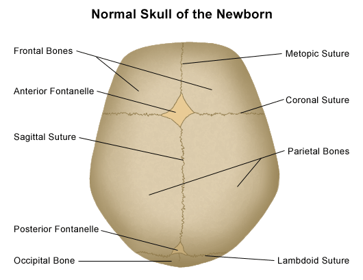
Cranial sutures are the fibrous joints that connect the bones of the skull. The primary sutures include:
- Sagittal suture - Between the two parietal bones.
- Coronal suture - Between the frontal bone and the parietal bones.
- Sphenoparietal suture - Between the sphenoid and parietal bones.
- Sphenosquamous suture - Between the sphenoid and squamous part of the temporal bone.
- Squamous suture - Between the parietal and temporal bones.
- Lambdoid suture - Between the parietal bones and the occipital bone.

Important Anatomical Landmarks
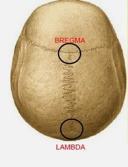
- Bregma: The point where the coronal suture intersects with the sagittal suture.
- Lambda: The point where the sagittal suture intersects with the lambdoid suture.

Neurocranium: Anterior View
Key anatomical structures visible from the anterior view of the neurocranium include:
- Frontal bone
- Superciliary arch
- Glabella
- Nasion
- Supra-orbital notch/foramen
- Zygomatic process
- Nasal bone
- Maxilla
- Frontal process
- Zygomatic process
- Infra-orbital foramen
- Alveolar process
- Anterior nasal spine
- Zygomatic bone
- Mandible
- Mental foramen
- Mental protuberance
- Mental tubercle
- Body, ramus, and angle of mandible
:background_color(FFFFFF):format(jpeg)/images/library/7258/Frontal_bone_01.png)
Neurocranium: Lateral View
Key sutures and anatomical structures visible from the lateral view:
- Sutures: Sphenosquamous, Coronal, Squamous, Sphenoparietal, Parietomastoid, Lambdoid, Occipitomastoid
- Bones:
- Parietal bone
- Frontal bone
- Nasal bone
- Lacrimal bone
- Zygomatic bone
- Maxilla
- Temporal bone
- Occipital bone
- Mandible
- Processes:
- Mastoid process
- Styloid process
- Zygomatic process of temporal bone
- Temporal process of zygomatic bone

Neurocranium: Posterior View
The posterior view of the skull highlights several important structures:
- Occipital bone
- External occipital protuberance
- External occipital crest
- Superior and inferior nuchal lines
- Occipital condyles
- Parietal bones
- Sagittal suture
- Lambdoid suture
- Parietal foramina
- Temporal bones
- Mastoid process
- Occipitomastoid suture
- Miscellaneous
- Sutural bones (Wormian bones) often found within the lambdoid suture area

Neurocranium: Inferior View
Structures visible from the inferior aspect of the neurocranium include:
- Maxilla and Palatine bones
- Hard palate (maxilla and palatine bones)
- Incisive fossa
- Greater and lesser palatine foramina
- Sphenoid bone
- Pterygoid processes (medial and lateral plates)
- Foramen ovale
- Foramen spinosum
- Temporal bone
- Mandibular fossa
- Carotid canal
- Jugular foramen
- Stylomastoid foramen
- Occipital bone
- Foramen magnum
- Occipital condyles
- Hypoglossal canal
- External occipital crest and protuberance
- Superior and inferior nuchal lines
Major Foramina and Their Contents
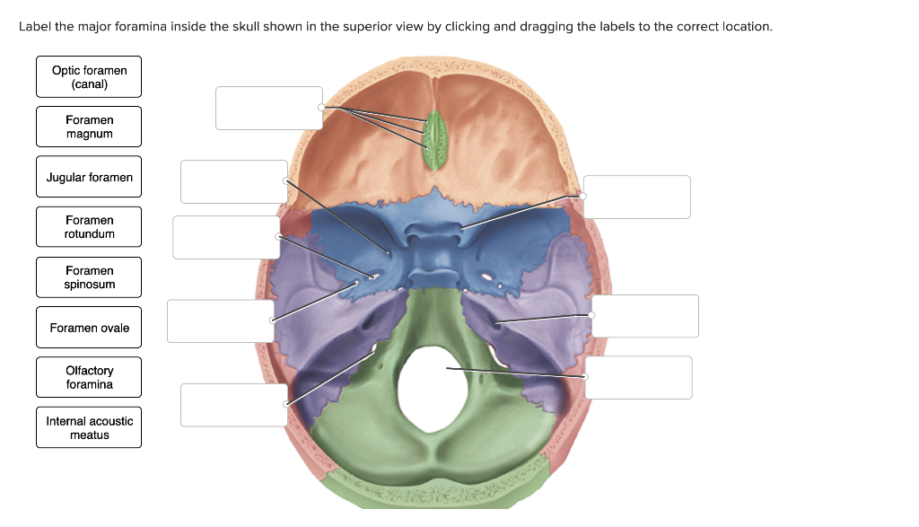
- Foramen ovale: Mandibular division of the trigeminal nerve (V3)
- Carotid canal: Internal carotid artery, lesser petrosal nerve
- Foramen spinosum: Middle meningeal artery, meningeal branch of mandibular nerve (V3)
- Hypoglossal canal: Hypoglossal nerve (XII)
- Jugular foramen: Glossopharyngeal nerve (IX), Vagus nerve (X), Accessory nerve (XI), internal jugular vein
- Foramen magnum: Spinal cord, vertebral arteries, roots of Accessory nerve (XI)
