Development of the Head, Neck, and Vertebral Column
Head and Neck Embryology
Summary Review of the 2nd Week of Development: 'Week of Two's'
- Two components of the trophoblast: Cytotrophoblast and Syncytiotrophoblast
- The embryoblast (inner cell mass) is converted to a bilaminar disc: Epiblast and Hypoblast
- Two new cavities form: Amnionic and Chorionic
- Two extraembryonic mesoderm layers: Somatopleuric and Splanchnopleuric

Summary Review of the 3rd Week of Development: 'Gastrulation, Neurulation, Cephalocaudal and Lateral Folding'
- Formation of the three embryonic germ layers
- Invaginating mesoderm cells form embryonic mesoderm
- The primitive streak and node are established in the epiblast
- The neural tube separates from the surface ectoderm
:background_color(FFFFFF):format(jpeg)/images/library/6092/Embryology_-_3rd_Week_of_Development__2_.png)
The Cranial Region of the Embryo Develops into the Head and Neck Beginning at the 4th Week
- Notable transformations during this period:
- Neural plate transforms via 'neurulation' to form neural folds and neural tube
- The formation of pharyngeal arches
- The development of sensory placodes: nasal, lens, and otic
- Limb buds and the stomodeum begin to form

Embryonic Derivatives in the Head and Neck
- From Ectoderm: Neural tube (CNS motor neurons, preganglionic ANS), neural crest (PNS postganglionic ANS), epithelial components of the skin, stomodeum, nasal pit, external auditory meatus, olfactory, lens, and otic placodes
- From Endoderm: Epithelial lining of the pharynx, larynx, trachea, esophagus, pharyngotympanic tube, middle ear; glands (thyroid, parathyroid, thymus, tonsils)
- From Mesoderm: Notochord, somites, somitomeres that form bone, cartilage, muscles, and connective tissues
The Neural Crest and Head Mesenchyme
- Neural crest cells give rise to the connective tissue and bones of the face and skull, cranial nerve ganglia, C cells of the thyroid gland, conotruncal septum of the heart, odontoblasts, dermis of the face and skin, spinal (dorsal root) ganglia, sympathetic and parasympathetic ganglia, adrenal medulla, Schwann cells, satellite cells, arachnoid and pia mater, and melanocytes
Pharyngeal Arches and Their Derivatives
During the 4th week:
- Develop as neural crest cells proliferate and migrate
- Support the lateral walls of the future pharynx.
- Initial appearance of 4 visible arches; the 6th arch is deeper
- Arch arteries coincide with aortic arches, forming key structures
Each pharyngeal arch has: - A cranial nerve supplying its epithelial, skeletal muscle, and connective tissue derivatives - Specific cartilage, muscle, and vascular derivatives, e.g.: - 1st Arch: Malleus, Incus - 2nd Arch: Stapes, Styloid process - 3rd Arch: Inferior hyoid - 4th and 6th Arches: Thyroid, cricoid, and other laryngeal cartilages
Development of the Face, Palate, and Tongue
- The first pharyngeal arch splits into maxillary and mandibular processes
- Frontonasal process forms around the forebrain
- Nasal placodes invaginate to form medial and lateral nasal swellings
- Intermaxillary segment forms the primary palate, philtrum of the upper lip, and the external nose. Maxillary processes form secondary palate.
- Tongue arises from pharyngeal arches:
- Anterior 2/3 from the 1st pharyngeal arch
- Posterior 1/3 from the 3rd and 4th pharyngeal arches
Somitomeres and Somites
- Organization of paraxial mesoderm begins around the third week
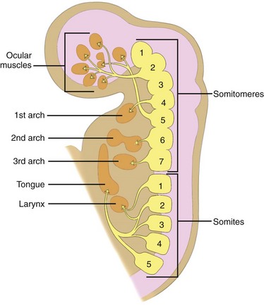
- In the head, forms 7 somitomeres which contribute mesenchyme
- In the rest of the body, organizes into somites, beginning in the occipital region and adding in a craniocaudal direction

Vertebral Development and Resegmentation
- Somite cells undergo 'epithelialization' and segment into sclerotome, myotome, and dermatome
- Sclerotomes migrate to form vertebrae and ribs
- During fourth week, sclerotomes resegment, so vertebrae consist of parts of adjacent sclerotomes
- Myotomes bridge vertebrae for spinal movement
- Failures in somitogenesis or resegmentation can lead to congenital scoliosis
Evaginations and Migration of Endoderm-Derived Structures
- Pharyngeal pouches form key glands: e.g., parathyroid, thymus
- Thyroid gland migrates from foramen cecum to final position
- Failure in migration or fusion can result in anatomical anomalies, like thyroglossal cysts or cleft lip/palate.
 ```
```