Protein Mutations and Diseases
Objectives
Explain how mutations can alter the properties or functions of proteins and produce specific diseases.

Protein mutations arise from changes in DNA
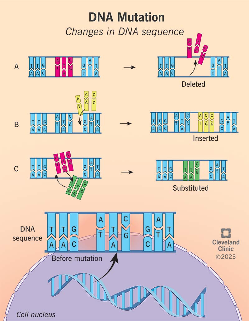
A permanent change in the nucleotide sequence of DNA is called a mutation. Mutations can involve the replacement of one base pair with another (substitution) or the addition or deletion of one or more base pairs (insertion or deletion). If the mutation affects non-essential DNA or has a negligible impact on the function of a gene product, it is called a silent mutation.
Mutations can arise: - from spontaneous chemical change in DNA - from exposure to chemicals/radiation - by defects in DNA/RNA metabolism
Mutations can be acquired in germ-line cells and passed on to offspring or acquired in somatic cells and cause disease such as cancer.

Types of mutations in DNA
Point mutation: change in a single base pair
- Transition point mutation: purine for purine (A for G or G for A) or pyrimidine for pyrimidine (T for C or C for T)
- Transversion point mutation: purine for pyrimidine or vice versa
- Point mutations further classified by effect:
- Missense mutation: the point mutation that changes a single base pair in a codon such that the codon now encodes a different amino acid.
- Nonsense mutation: the point mutation that changes a single base pair in a codon to a stop codon that terminates translation.
- Silent mutation: does not alter the amino acid encoded.
Frameshifts: Additions or deletions of one or more bases (if not a multiple of 3) will lead to frameshifts that disrupt the coding of a protein.
Mutations can alter the structure/function of proteins
- Missense mutation: An incorrect amino acid, which may produce a malfunctioning protein.
- Nonsense mutation: Incorrect sequence causes shortening of the protein.
Insertion and Deletion Mutations
- Insertion mutation: Incorrect amino acid sequence, which may produce a malfunctioning protein due to the insertion of a single nucleotide.
- Deletion mutation: Incorrect amino acid sequence, which may produce a malfunctioning protein due to the deletion of a single nucleotide.

Repeat Expansion Mutation
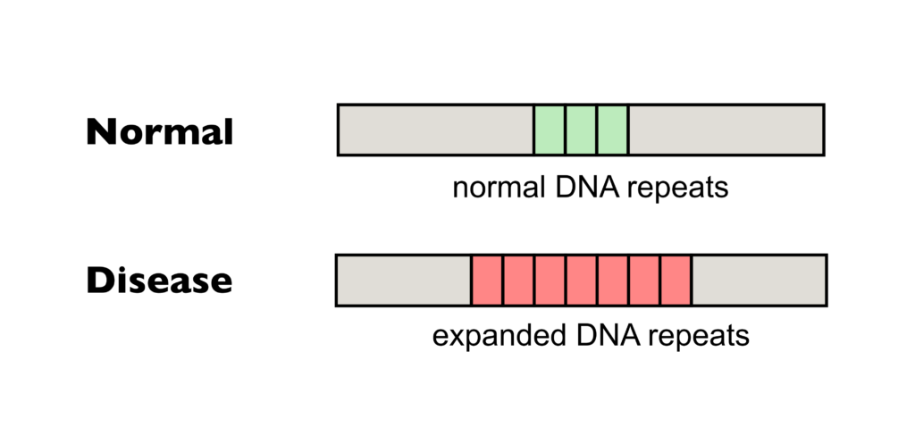
A repeated trinucleotide sequence (CAG) adds a string of glutamines (Gln) to the protein.
Genetic Disorders - Four main types
- Single gene (Mendelian or monogenic): Caused by changes or mutations that occur in the DNA sequence of one gene. Examples include cystic fibrosis, sickle cell anemia, Marfan syndrome, and Huntington’s disease.
- Multifactorial (complex or polygenic): Caused by a combination of environmental factors and mutations in multiple genes. Examples include heart disease, high blood pressure, arthritis, diabetes, cancer, and obesity.
- Chromosomal: Abnormalities in chromosome structure, such as missing or extra copies or gross breaks and rejoinings (translocations). Examples include Down syndrome or trisomy 21 and certain types of leukemia.
- Mitochondrial diseases: Caused by mutations that impact mitochondrial function. Examples include Leber’s hereditary optic neuropathy, MELAS syndrome, and Kearns-Sayre syndrome.
Examples of mutations that affect protein structure and function and cause specific diseases
- Hemoglobinopathies: More than 1,000 mutations in the genes encoding the α- and β-globin polypeptides have been documented. Types include unstable hemoglobins and those with increased oxygen affinity.
- p53 Tumor Suppressor Gene (Cancer): Mutations prevent p53 from binding DNA. This form of mutation is termed dominant negative.
- Cystic Fibrosis: The most common mutation results in defective processing and trafficking of the protein, leading to improper chloride channel activity in the affected cell.

Hemoglobinopathies
Hemoglobinopathies include all genetic hemoglobin disorders, divided into structural hemoglobin variants and thalassemia syndromes. For example, sickle cell disease is caused by a missense mutation that results in the substitution of valine for glutamate at position 6 on the β-subunit of hemoglobin.
p53 Tumor Suppressor Gene
The majority of p53 mutations in cancer are in the DNA binding domain. These mutations prevent p53 from binding to promoters of its target genes in response to DNA damage, leading to survival and possible replication of cells with a corrupted genome.
Cystic Fibrosis
Cystic Fibrosis is characterized by a defect in CFTR gene leading to abnormal chloride ion transport. The most common mutation is the deletion of phenylalanine at position 508 (F508del), resulting in improper protein folding and function.