Anatomy of the Pelvic Girdle and Perineum
Anatomy of the Pelvic Girdle and Perineum
Pelvic Girdle Bones
The pelvic girdle comprises several bones intricately joined to provide structural stability and support for various body functions. Here's a closer look at the bones of the pelvic girdle:
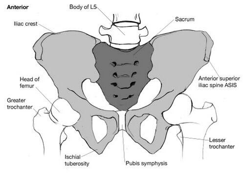
- Iliac Crest: The curved superior border of the ilium, forming the prominent ridge on the top of the hip bone.

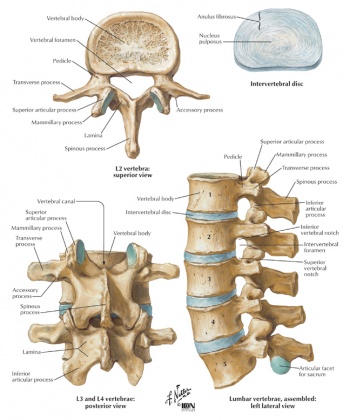
- L5 Vertebra: The fifth lumbar vertebra, key in transferring weight from the upper body to the pelvis.
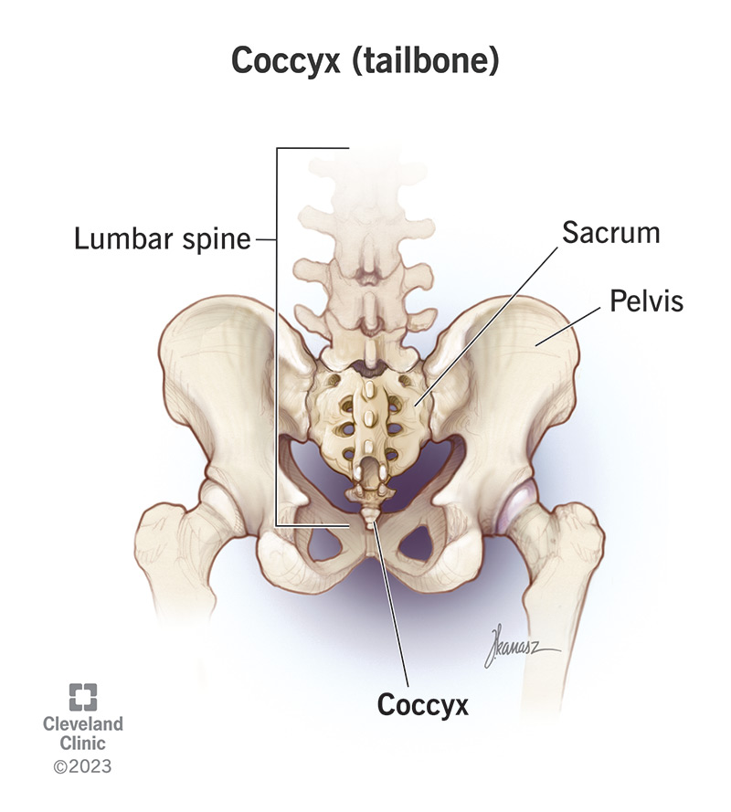
- Sacrum: A triangular bone at the base of the spine, forming the posterior section of the pelvis.
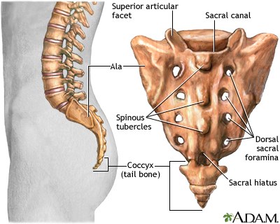
- Iliac Fossa: A large, smooth, concave surface on the internal side of the ilium, supports the iliacus muscle.

- Ala of Sacrum: The wide, wing-like edges of the sacrum, engaging in the sacroiliac joint.
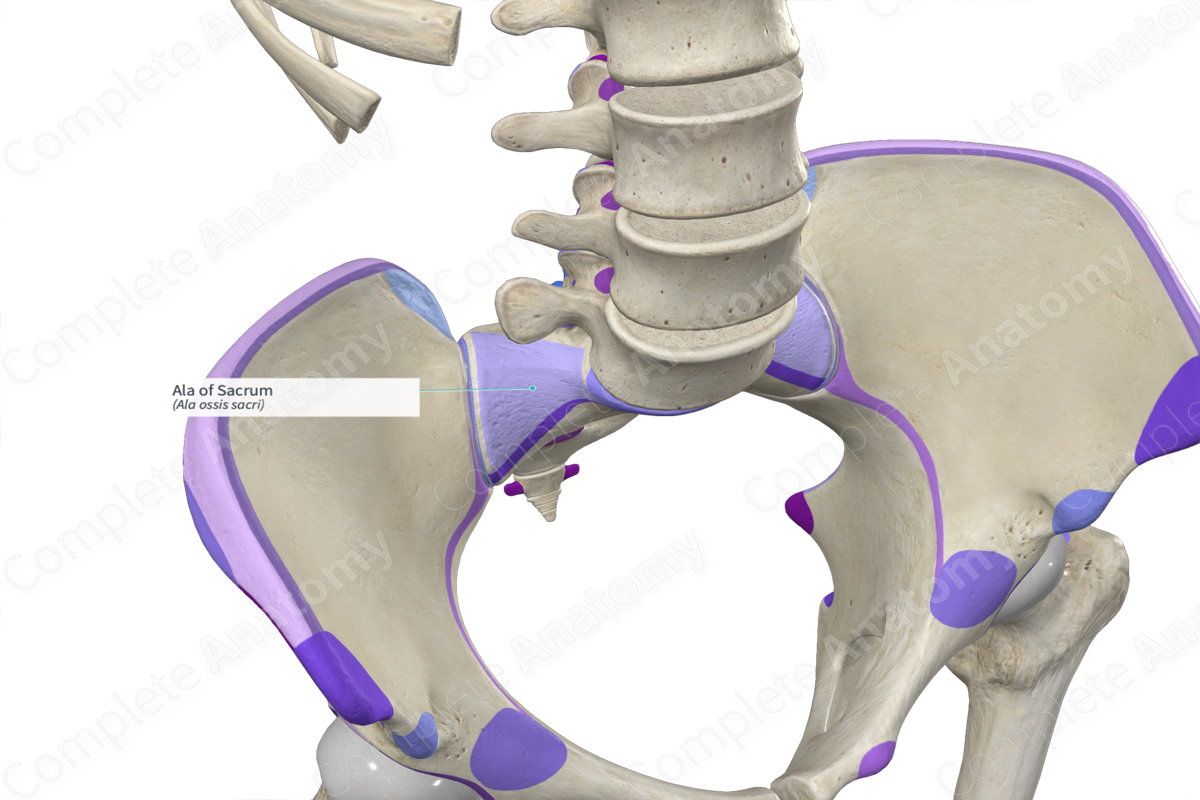
- Anterior Superior Iliac Spine: A palpable bony projection at the anterior end of the iliac crest.
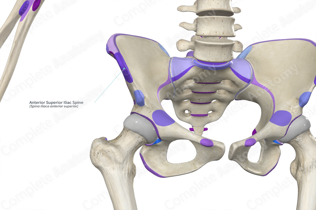
- Sacral Promontory: The anteriorly projecting edge of the sacral base, marking the pelvic inlet.
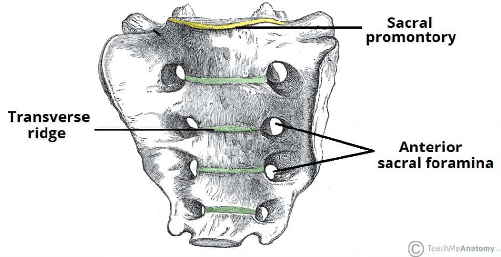
- Anterior Sacral Foramina: Openings in the sacrum that allow for the passage of nerves and blood vessels.

- Anterior Inferior Iliac Spine: A projection below the anterior superior iliac spine, serving as an attachment point for muscle.
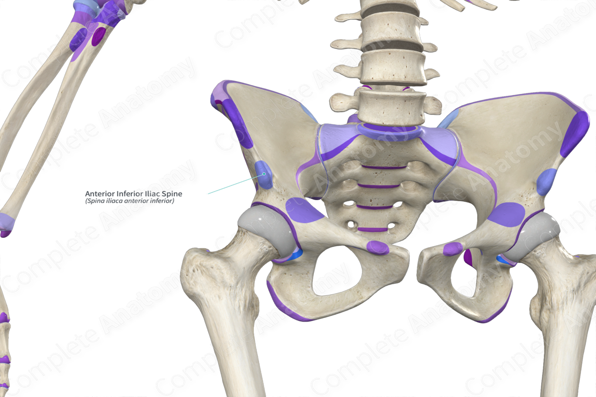
- Iliopectineal Line: A ridge running along the superior pubic ramus to the arcuate line, important in demarcating the pelvis.
- Arcuate Line: Part of the pelvic brim, continuous with the iliopectineal line.

- Ischial Spine: A bony projection from the ischium, separating the greater and lesser sciatic notches.
:background_color(FFFFFF):format(jpeg)/images/article/ischial-spine/mOxQpiZVpA5GtbVCNoCPQ_MUY7m3J7l62a95Nc8pmXCQ_Ischial_spine.png)
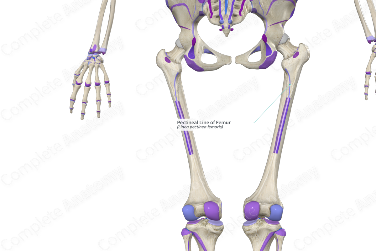
- Pectineal Line: A ridge on the superior ramus of the pubis.
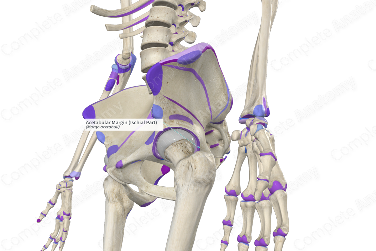
- Acetabular Margin: The rim of the acetabulum, articulating with the head of the femur.
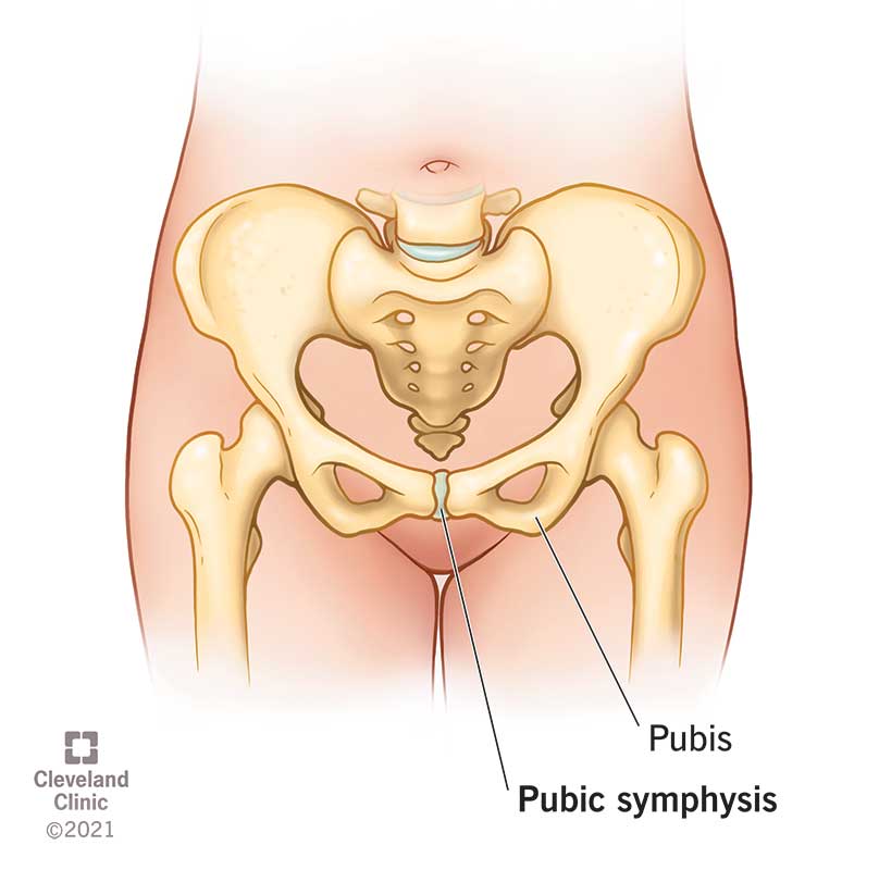
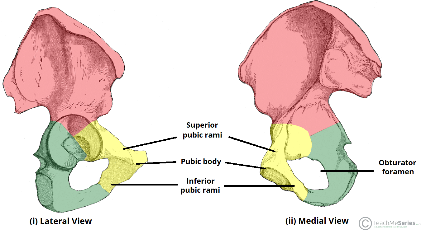
- Superior Pubic Ramus: The upper segment of the pubic bone.
- Pubic Tubercle: A prominent tubercle on the superior border of the pubic bone.
:background_color(FFFFFF):format(jpeg)/images/article/pubic-tubercle/oMRfXj5ygPgGy8WOQq3PAw_Pubic_tubercles.png)
- Pubic Crest: The thick anterior part of the bone that connects the pubic tubercles.
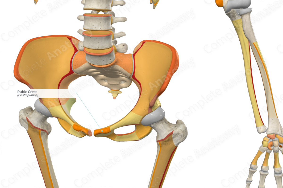
- Inferior Pubic Ramus: The lower part of the pubic bone, joining the ischial ramus.

- Ischial Ramus: Part of the ischium, contributing to the obturator foramen.

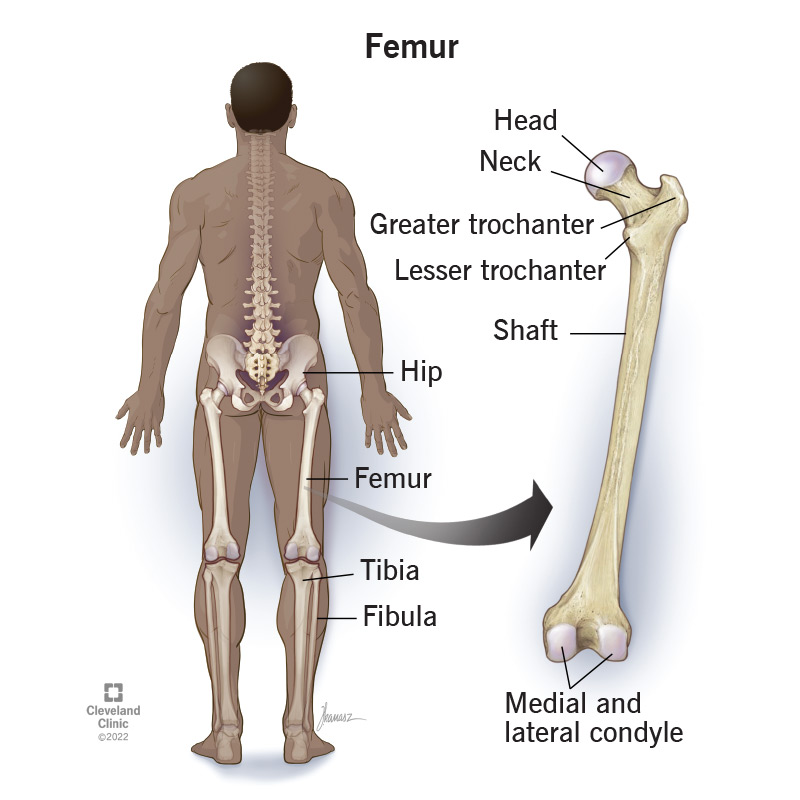
- Femur: The thigh bone, fitting into the acetabulum to form the hip joint.
- Coccyx: The terminal part of the vertebral column.
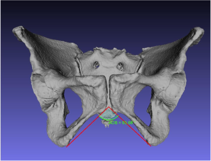
- Subpubic Angle: The angle formed below the pubic symphysis, more acute in males.
- Obturator Foramen: The large opening in the hip bone, surrounded by the pelvis.

- Pubic Symphysis: A cartilaginous joint uniting the left and right pubic bones.
Surface Anatomy
Landmarks (Anterior View)
- L5 Vertebra, Iliac Crest, Iliac Tubercle: Key landmarks palpable on the body.
- Gluteal Lines (Posterior, Anterior, Inferior): Indicate muscle attachment sites on the ilium.

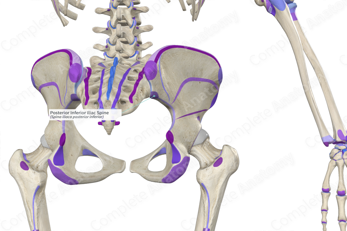
- Posterior Superior Iliac Spine, Posterior Inferior Iliac Spine: Bony landmarks used in clinical examinations.
- Sacral Hiatus, Ischial Spine, Pubic Symphysis: Critical anatomical points related to nerve and vascular structures.

Pelvic Cavity
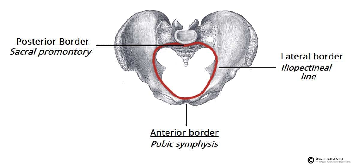
- Pelvic Inlet (Pelvic Brim): The entrance to the true pelvis, demarcated by the linea terminalis.
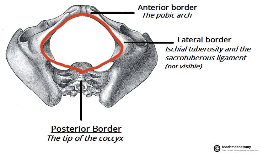
- Pelvic Outlet: The exit of the true pelvis, bordered by the pubic arch and sacrum.
Sexual Dimorphism of the Pelvis
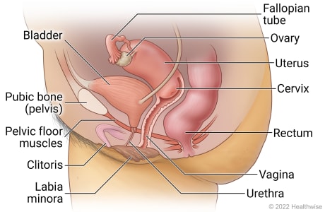
- Female Pelvis: Wider and shallower, with a larger pelvic inlet and outlet to facilitate childbirth.
- Male Pelvis: Narrower and deeper, with prominent bony landmarks.
:watermark(/images/watermark_5000_10percent.png,0,0,0):watermark(/images/logo_url.png,-10,-10,0):format(jpeg)/images/overview_image/262/PrGv4z1Vu7AoPiRrD1hnRw_male-pelvic-viscera-and-perineum_english.jpg)
Measurements
- Subpubic Angle: Wider in females (80º-85º) compared to males (50º-60º).

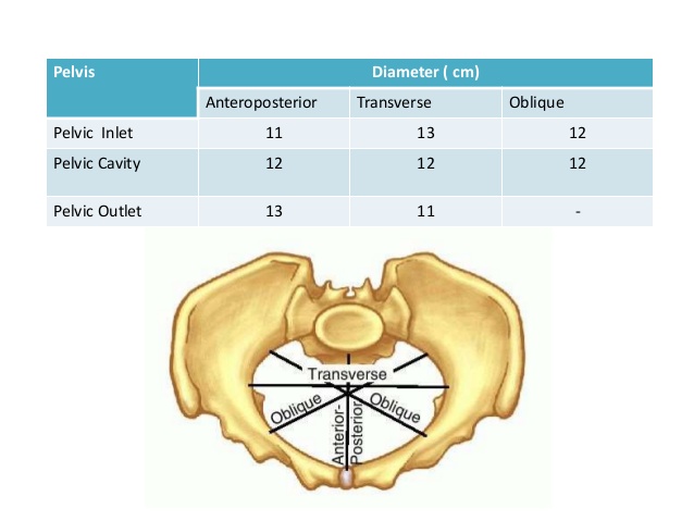
- Conjugate Diameter of Pelvic Inlet: Approximately 11 cm.

- Transverse Diameter of Pelvic Inlet: Around 13 cm.
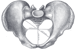
- Anteroposterior Diameter of Pelvic Outlet: Ranges from 9.5 to 11.5 cm.
Lesser and Greater Pelvis
- True (Lesser) Pelvis: Contains the pelvic organs.
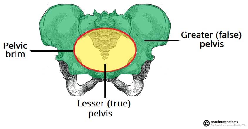
- False (Greater) Pelvis: Part of the abdominal cavity, superior to the pelvic brim.
Pelvic Diaphragm
- Structure: A bowl-shaped group of muscles including the levator ani and coccygeus muscles, supporting pelvic organs and maintaining continence.
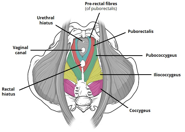
- Openings: Allow passage for gastrointestinal and genitourinary systems.

The Anal and Urogenital Triangles
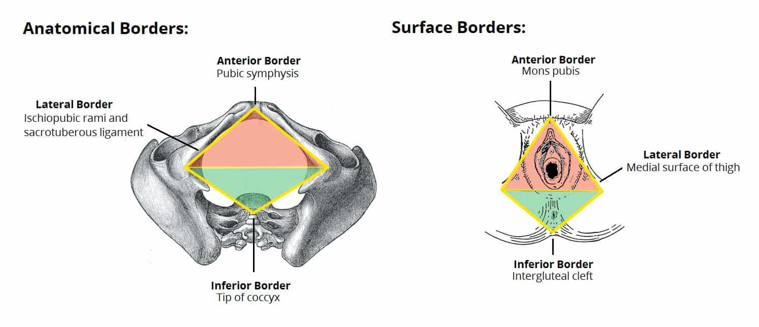
- Components: Anterior urogenital triangle and posterior anal triangle.

- Boundaries: Defined by bony landmarks: pubic symphysis, ischial tuberosities, and coccyx.
Perineal Muscles and Pouches
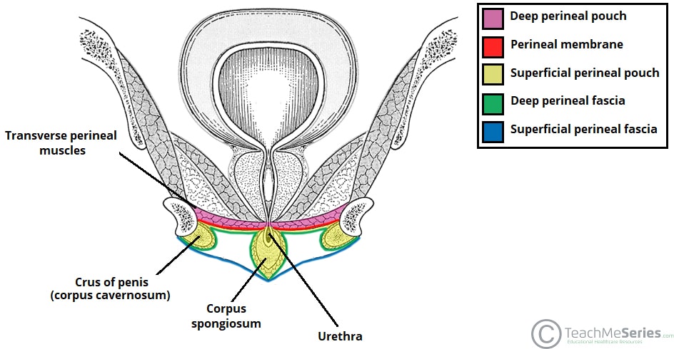
Male Perineum
- Superficial and Deep Perineal Pouch: Contain muscles and structures vital for urinary and sexual functions.
Female Perineum
- Bulb of Vestibule, Greater Vestibular Glands: Erectile tissues and glands essential for reproductive health.

Summary
Understanding the detailed structure and anatomy of the pelvic girdle and perineum is crucial for recognizing the functional implications and clinical significance of this region. This comprehensive overview covers the essential bones, muscles, and anatomical landmarks defining the pelvic region, essential for medical and anatomical studies. :watermark(/images/watermark_5000_10percent.png,0,0,0):watermark(/images/logo_url.png,-10,-10,0):format(jpeg)/images/overview_image/262/PrGv4z1Vu7AoPiRrD1hnRw_male-pelvic-viscera-and-perineum_english.jpg)
