Cranial Nerves: Structure, Function, and Reflexes
Cranial Nerves Overview
CN III: Oculomotor Nerve
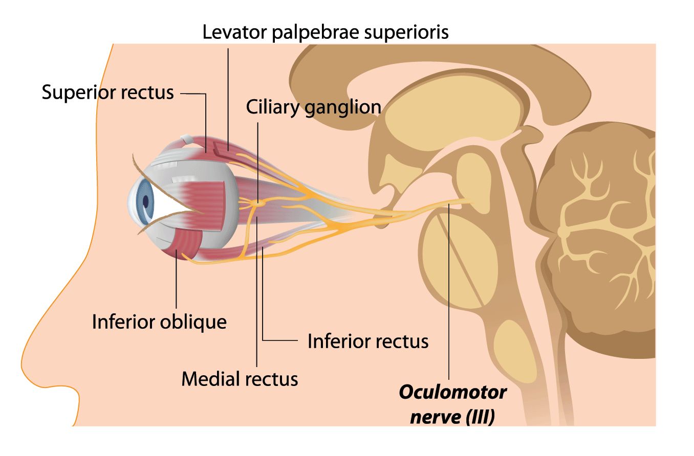
The oculomotor nerve (CN III) travels through the superior orbital fissure and carries preganglionic parasympathetic fibers that innervate the pupillary sphincter (causing pupillary constriction) and the ciliary muscles (for lens accommodation). These fibers originate in the midbrain and synapse at the ciliary ganglion.
CN VII: Facial Nerve
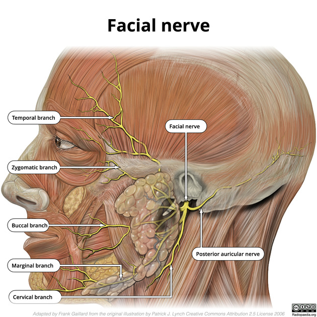
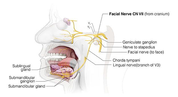
The facial nerve (CN VII) exits the skull via the internal acoustic meatus. This nerve has several branches:
- Facial nerve proper: Innervates muscles of facial expression and stapedius muscle, which dampens sound.
- Greater petrosal nerve: Provides lacrimation by innervating the lacrimal gland and nasal glands (via the pterygopalatine ganglion).
:max_bytes(150000):strip_icc()/greaterpetrosalnerve-fd0e279bf0f343b59b01dea1b722cc8d.jpg)
- Chorda tympani: Controls salivation by innervating the submandibular and sublingual glands. It travels with the lingual nerve (branch of V3) and carries taste fibers from the anterior 2/3 of the tongue.
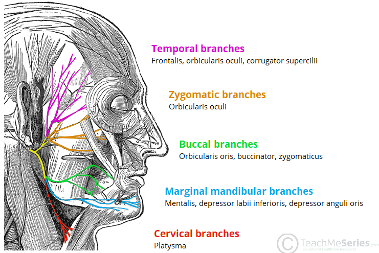
Branches of Facial Nerve Proper
- Temporal branches: Innervate frontalis, orbicularis oculi, and corrugator supercilii.
- Zygomatic branches: Innervate orbicularis oris.
- Buccal branches: Innervate orbicularis oris, buccinator, and zygomaticus.
- Marginal mandibular branches: Innervate mentalis, depressor labii inferioris, and depressor anguli oris.
- Cervical branches: Innervate platysma.
CN IX: Glossopharyngeal Nerve
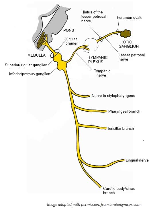
The glossopharyngeal nerve (CN IX) exits the skull via the jugular foramen. It provides:
- Motor innervation to the stylopharyngeus muscle.
- General sensation to the tympanic membrane.
- Taste and general sensation to the posterior 1/3 of the tongue.

Branches of CN IX
- Lesser petrosal nerve: Controls salivation by innervating the parotid gland. This nerve travels closely with the middle meningeal artery and synapses at the otic ganglion.
- Auriculotemporal nerve: Carries sensation from the ear, temporomandibular joint (TMJ), and the area around the Eustachian tube.
CN V: Trigeminal Nerve
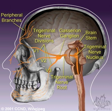
The trigeminal nerve (CN V) has three main branches:
- Ophthalmic nerve (V1): Sensory only. Passes through the superior orbital fissure and supplies sensation to the eye (cornea and sclera), forehead, and upper eyelid.
- Maxillary nerve (V2): Sensory only. Passes through the foramen rotundum and supplies sensation to the middle part of the face (including the upper teeth and palate).
- Mandibular nerve (V3): Sensory and motor. Passes through the foramen ovale and supplies sensation to the lower part of the face (including lower teeth and anterior 2/3 of the tongue) and motor innervation to muscles of mastication and the tensor tympani muscle.
Detailed Branches of Trigeminal Nerve
- Ophthalmic nerve (V1): Includes nasociliary, frontal, supraorbital, and lacrimal nerves.
- Maxillary nerve (V2): Includes zygomatic, infraorbital, nasopalatine, superior alveolar, and greater/lesser palatine nerves.
- Mandibular nerve (V3): Includes buccal, inferior alveolar, auriculotemporal, and lingual nerves, and motor branches to muscles of mastication and the tensor tympani muscle.
Cranial Nerve Reflexes
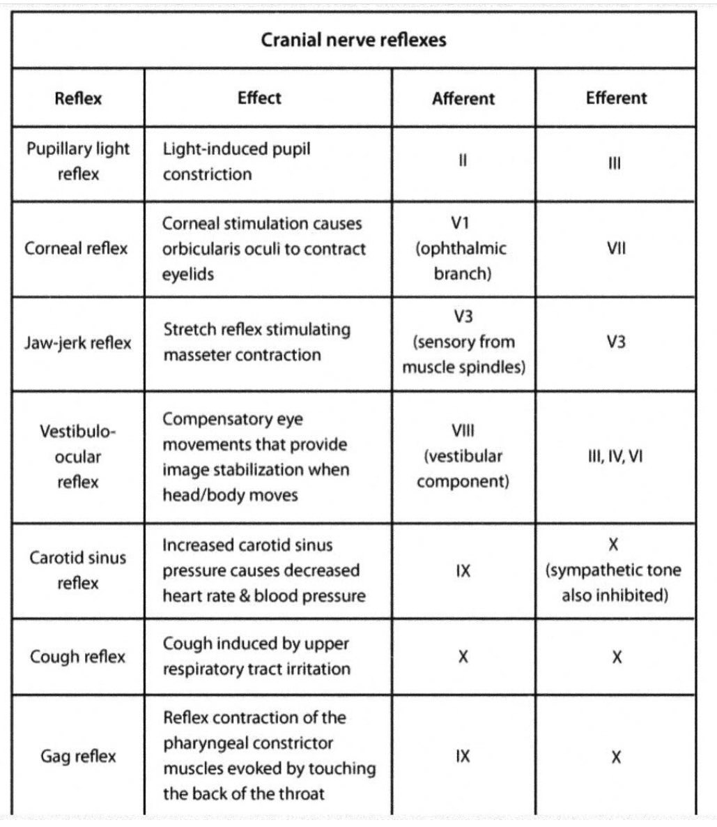
- Corneal Reflex (Blinking):
- Afferent: Nasociliary nerve of the ophthalmic branch (V1).
- Efferent: Temporal and zygomatic branches of the facial nerve (CN VII).
- Pupillary Reflex:
- Afferent: Optic nerve (CN II).
- Efferent: Oculomotor nerve (CN III).
- Gag Reflex:
- Afferent: Glossopharyngeal nerve (CN IX).
- Efferent: Vagus nerve (CN X).