Formation of Trilaminar Germ Disc and Differentiation of Germ Layers
Formation of Trilaminar Germ Disc and Germ Layer Differentiation
Formation of Trilaminar Germ Disc (3rd Week)
The formation of the trilaminar germ disc occurs during the third week of embryonic development. This transformation is pivotal as it transitions the embryo from a bilaminar disc, comprised of the epiblast and hypoblast, to a trilaminar disc composed of three germ layers: ectoderm, mesoderm, and endoderm. This process is known as gastrulation and is initiated with the appearance of the primitive streak.
Appearance of the Primitive Streak
The primitive streak appears as a thickened linear band on the caudal surface of the epiblast. It is a critical structure that establishes the embryo's craniocaudal axis. At the cranial end of the streak lies an elevated area called the primitive node, which surrounds a small depression known as the primitive pit. 
Proliferation and Migration of Epiblast Cells
Epiblast cells proliferate and migrate through the primitive streak, a process regulated by fibroblast growth factor (FGF8). FGF8 downregulates E-cadherin expression, facilitating the movement of these cells. This migration converts the bilaminar disc into a trilaminar disc composed of the ectoderm, mesoderm, and endoderm. 
Formation of the Three Germ Layers
- Ectoderm: Cells that remain in the epiblast form the ectoderm.

- Mesoderm: Migrating cells that position themselves between the epiblast and hypoblast layers form the mesoderm.

- Endoderm: Some migrating cells displace the hypoblast cells to form the endoderm.

Widespread Migration of Mesenchymal Cells
Mesenchymal cells derived from the epiblast spread out to eventually form a complete layer of mesoderm. This layer connects to the extraembryonic mesoderm, contributing to the formation of structures such as the somatopleuric and splanchnopleuric layers.
Germ Layer Derivatives and Neurulation
Derivatives of the Germ Layers
- Ectoderm: Gives rise to the central and peripheral nervous systems, sensory epithelium of the ear, nose, and eye, epidermis, hair, nails, and glands.

- Mesoderm: Develops into muscle, cartilage, bone, connective tissue, subcutaneous skin tissues, the cardiovascular system, and the urogenital system.

- Endoderm: Forms the epithelial lining of the gastrointestinal and respiratory tracts, urinary bladder, and the parenchyma of glands like the tonsils, thyroid, parathyroid, thymus, liver, and pancreas.

Neurulation
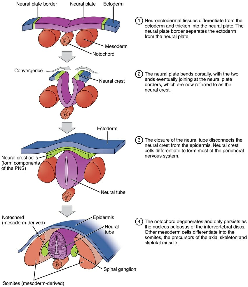
Neurulation starts with the notochord's induction of the overlying ectoderm to form the neural plate. The neural plate's lateral edges elevate to create neural folds, which will fuse to form the neural tube. This tube eventually separates from the surface ectoderm, forming the central nervous system. 
Neural Crest Differentiation
Neural crest cells undergo an epithelial-mesenchymal transition and migrate to various parts of the embryo. These cells give rise to several structures, including spinal and cranial nerve ganglia, adrenal medulla, autonomic ganglia, glial cells, Schwann cells, and melanocytes. 
Ectoderm, Mesoderm, and Endoderm Differentiation
- Ectoderm: Forms the epidermis and neural tissues.

- Mesoderm: Differentiates into paraxial mesoderm (forming somites), intermediate mesoderm (forming urogenital structures), and lateral plate mesoderm, which splits into two layers to form the intraembryonic coelom.

- Endoderm: Develops into the epithelial linings of internal organs and glands.

Cephalocaudal and Lateral Folding
Embryonic disc's rapid growth, along with cephalocaudal and lateral folding, shapes the embryo into a cylindrical form. This folding results in the constriction of the yolk sac and its partial incorporation into the embryo, forming the primitive gut. 