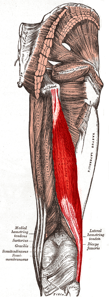
Posterior Thigh and Popliteal Fossa
Posterior Thigh and Popliteal Fossa
Anatomy of the Posterior Thigh and Popliteal Fossa
Bones
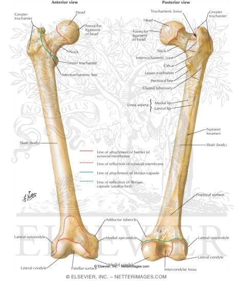
Femur
- Head
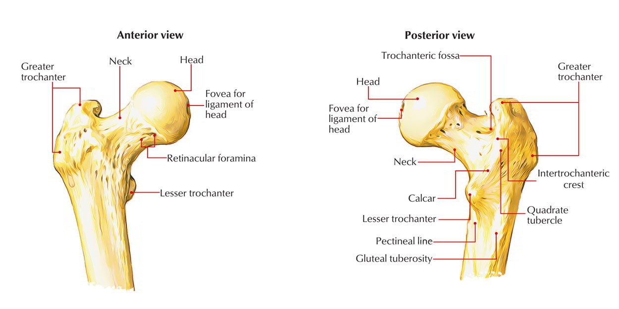
- Linea Aspera
- Lateral Supra Condylar Line
- Medial Supra Condylar Line

- Popliteal Surface
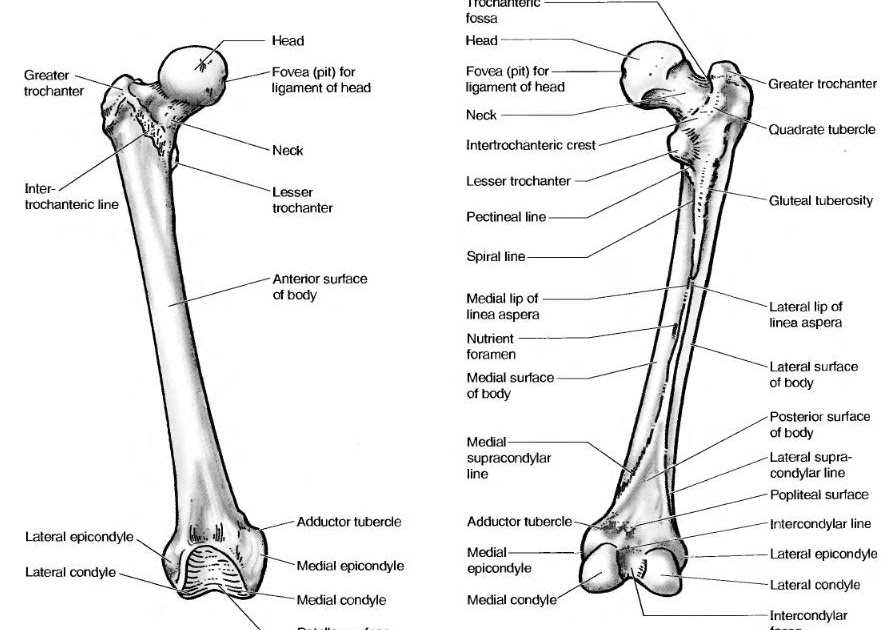
- Medial Condyle
- Intercondylar Fossa

- Lateral Condyle
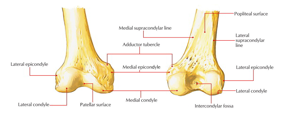
- Netter 489
Tibia & Fibula
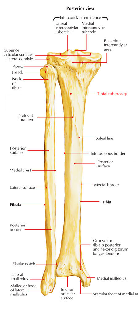
- Intercondylar Eminence
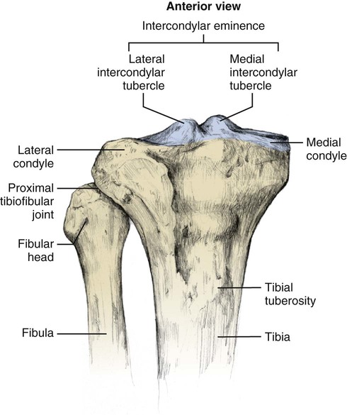
- Medial Condyle
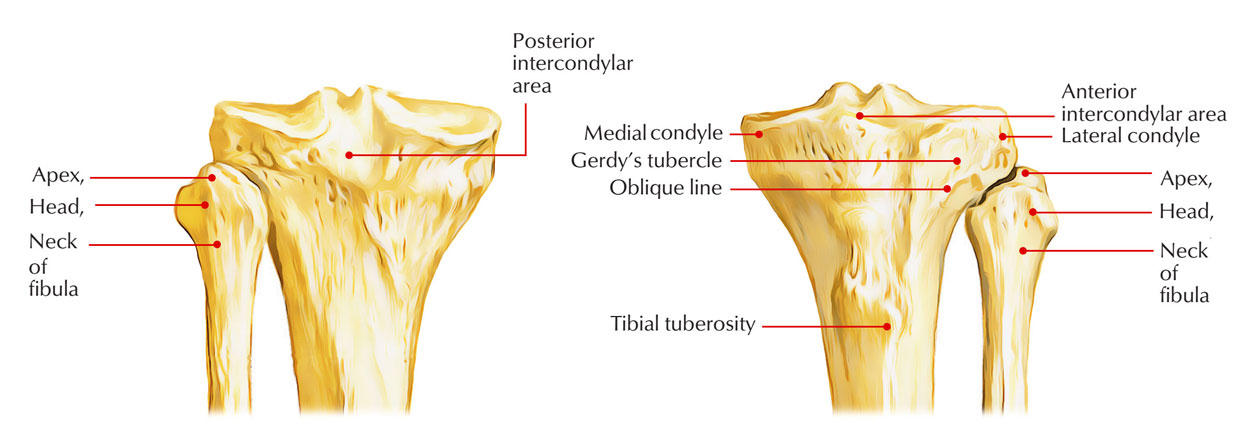
- Lateral Condyle

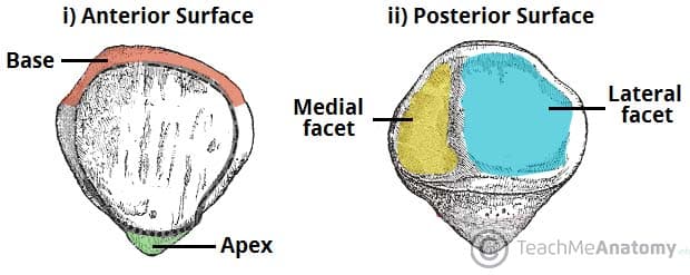
- Apex
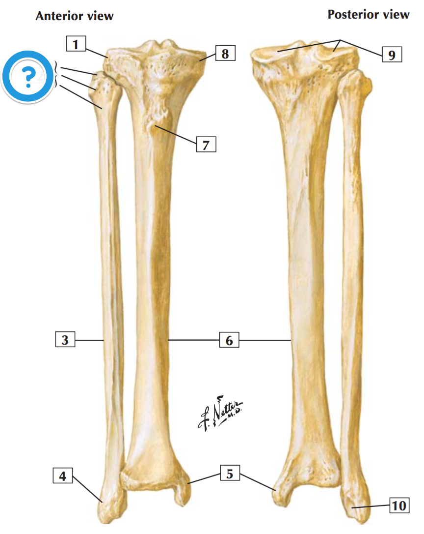
- Head of Fibula

- Neck

- Soleal Line
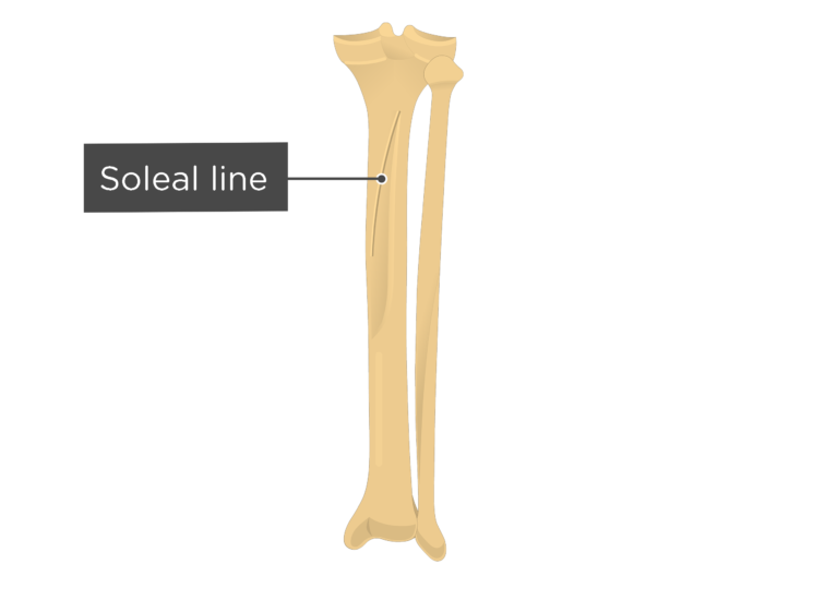
- Tibia: Transmits the body weight from the femur to the talus.
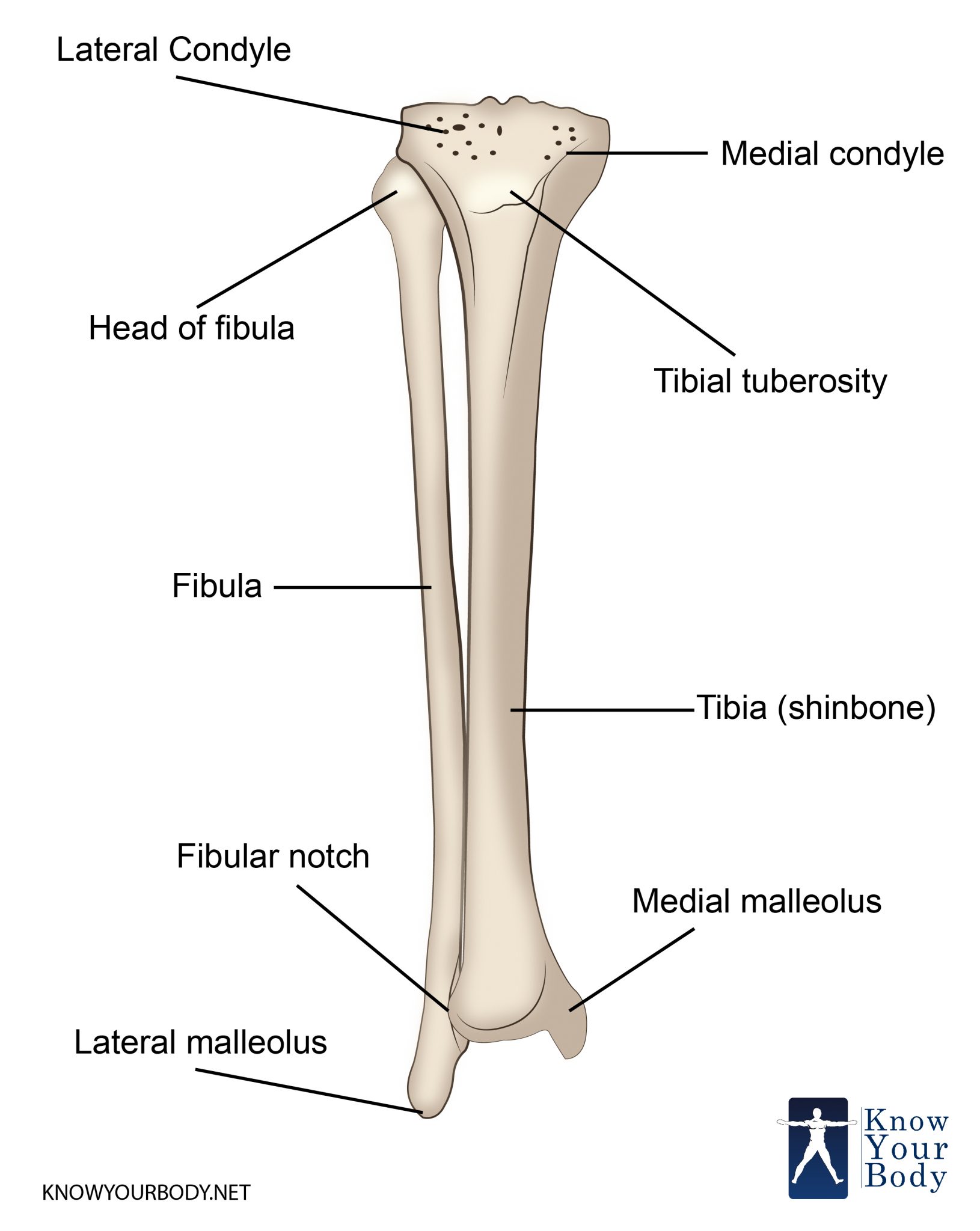
- Fibula: Has no weight-bearing function. (Netter 513)
Knee Joint
Sagittal Section of Knee Joint
- Femur
- Knee Joint:
- Quadriceps Femoris Tendon

- 2 femorotibial articulations

- 1 femoropatellar articulation
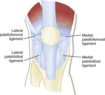
- Quadriceps Femoris Tendon
- Suprapatellar Bursa

- Patella
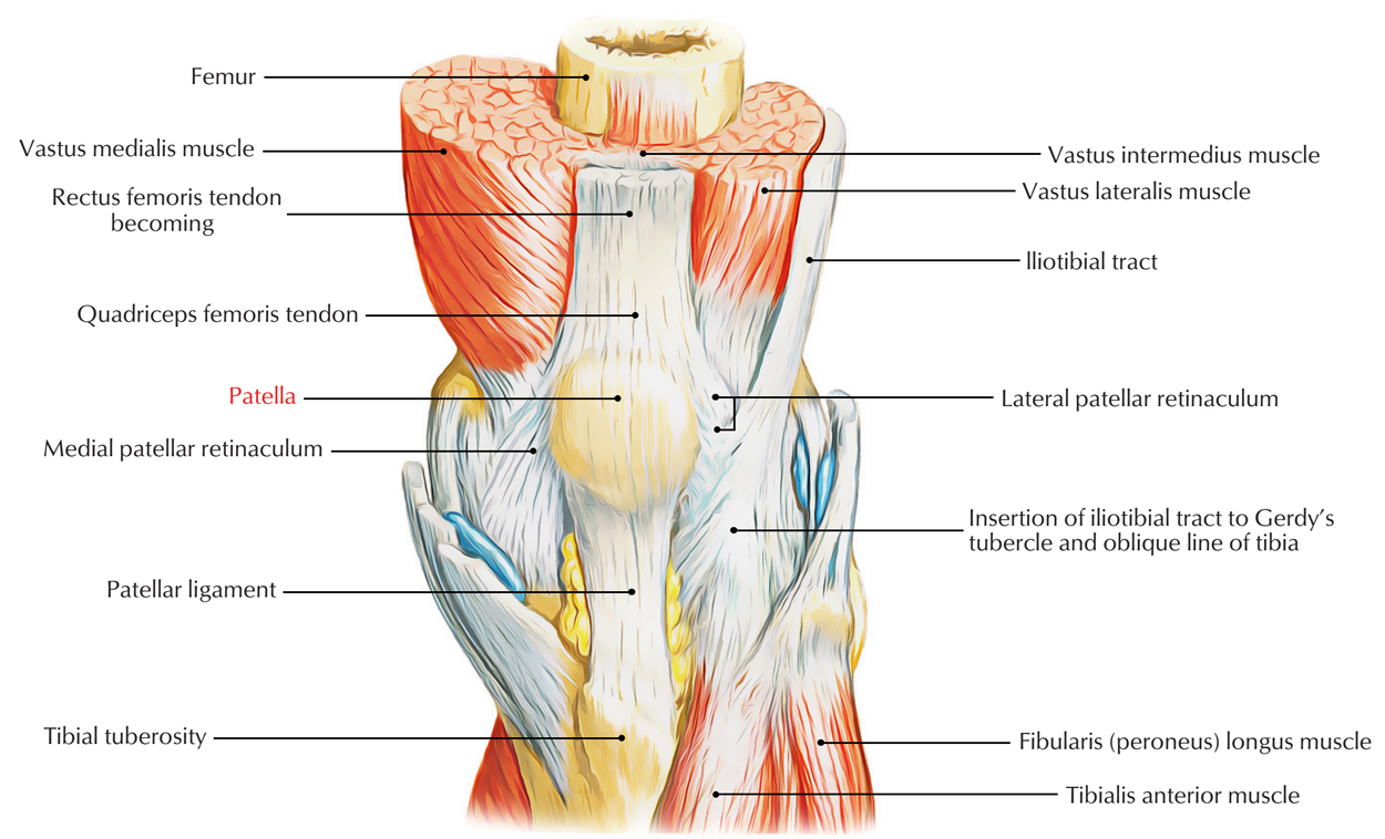
- Joint Capsule

- Synovial Membrane
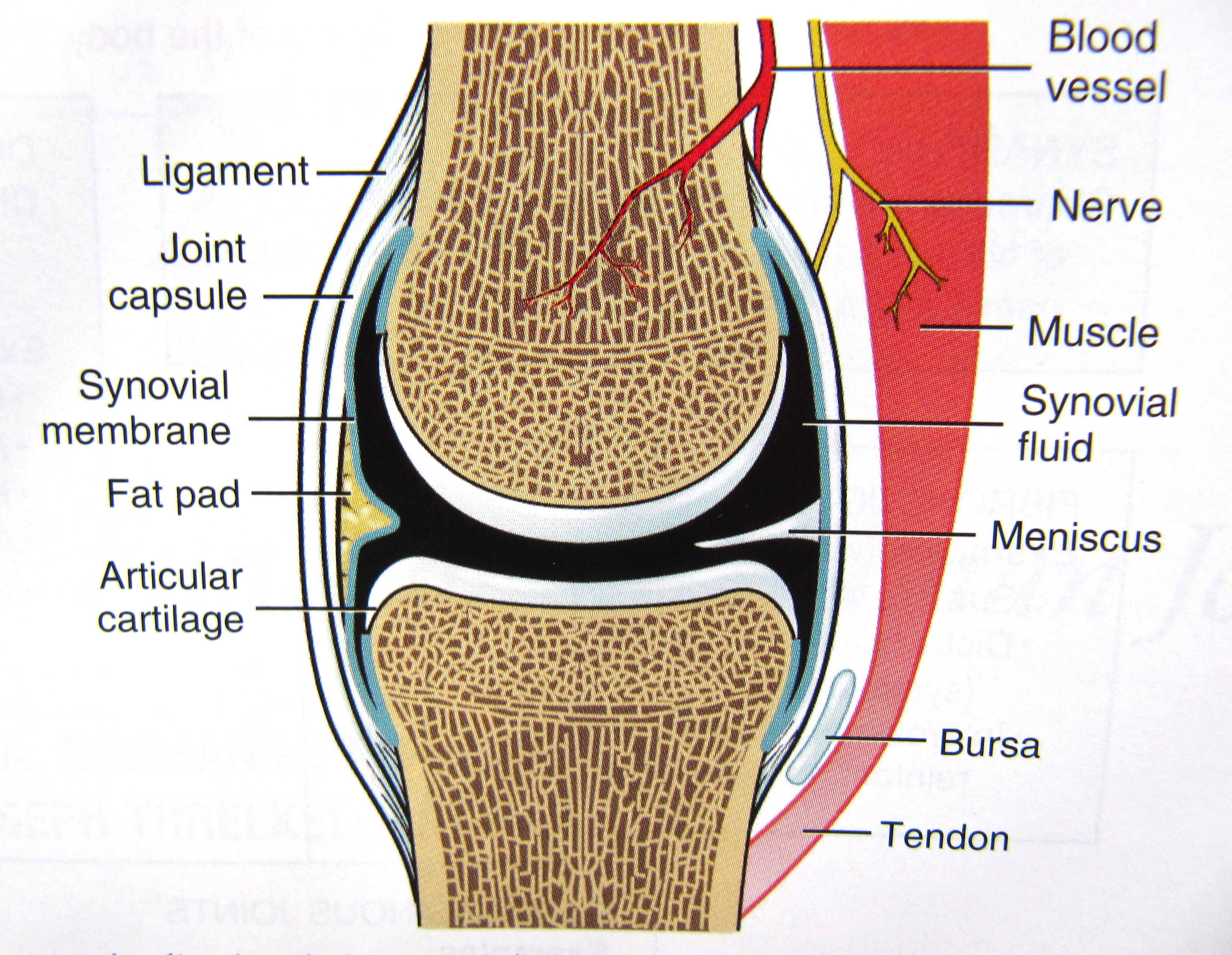
- Articular Cartilage

- Infrapatellar Fat Pad

- Patellar Ligament

- Fibula is not involved in the knee joint.
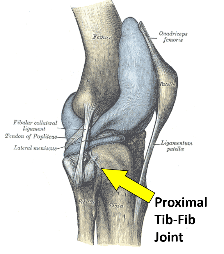
- Tibial Tuberosity
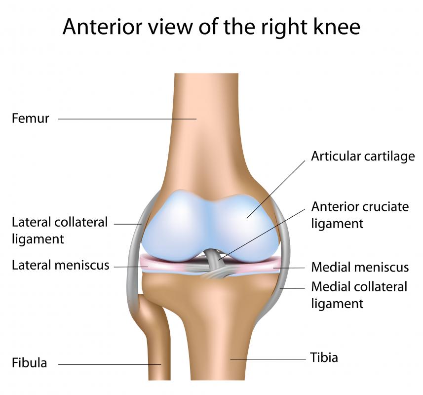
Knee Joint: Anterior View
- Lateral Condyle of Femur

- Medial Condyle of Femur

- Infrapatellar

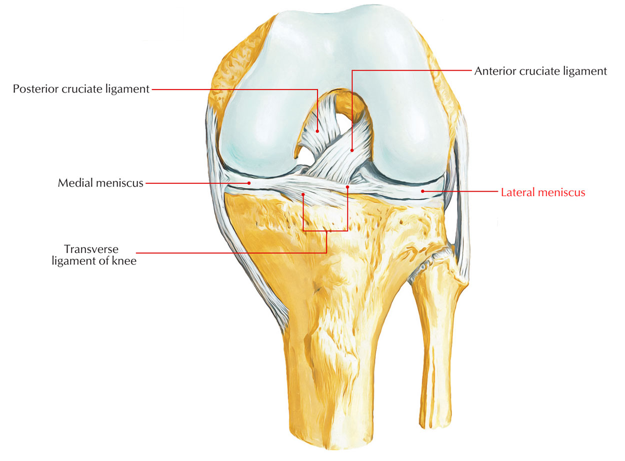
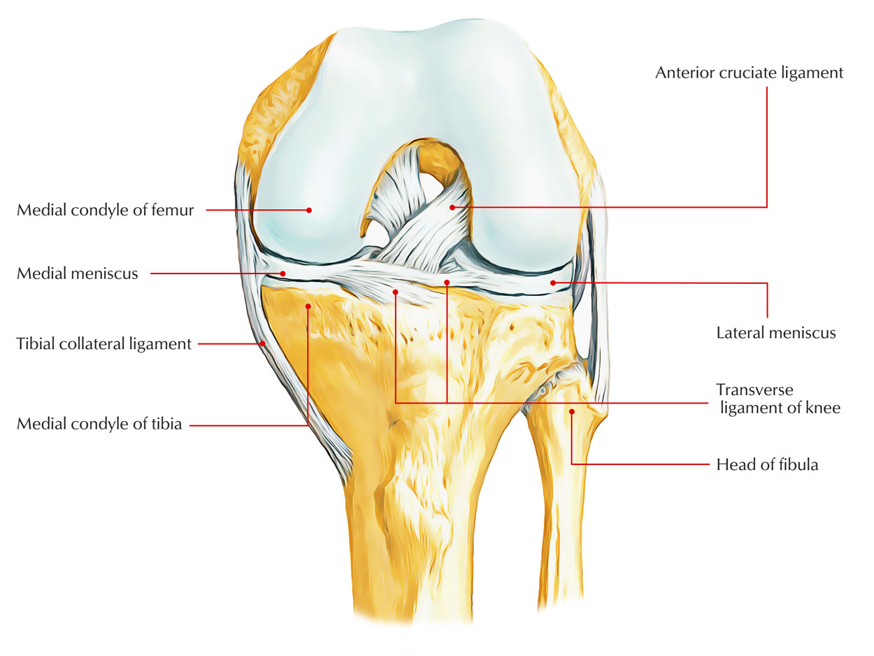
- Lateral Meniscus
- Synovial Fold

- Medial Meniscus
- Infrapatellar Pad of Fat

- Patella
- Suprapatellar Bursa: The superior extension of the joint cavity forms the suprapatellar bursa.

Knee Joint (Posterior View): Extracapsular Ligaments
- Attachment of Joint Capsule:
- Attached to the medial femur = medial condyle of the femur.
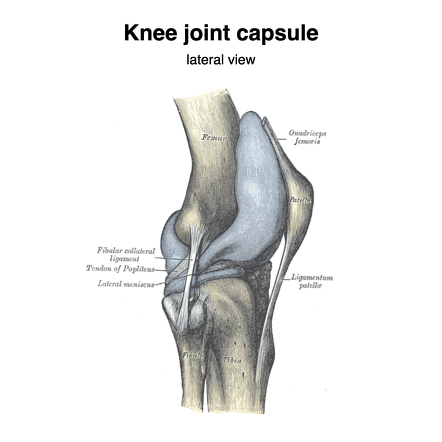
- Among others, the tibial collateral and fibular collateral ligaments provide additional support.

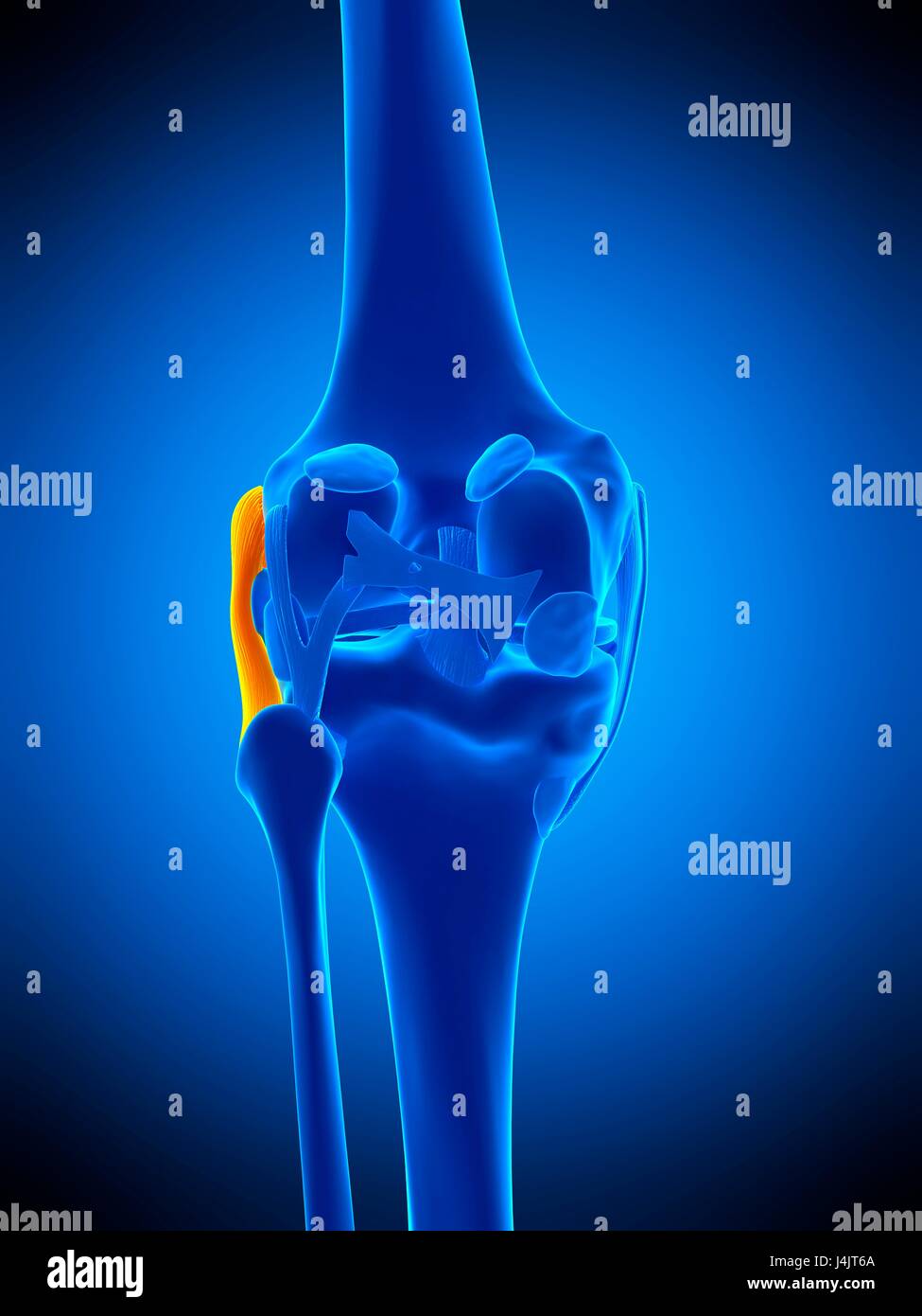
- Attached to the medial femur = medial condyle of the femur.
Cruciate Ligaments and Menisci
Intra-articular Ligaments
- Anterior Cruciate Ligament (ACL): Limits the posterior rolling of the femoral condyles on the tibia during flexion.
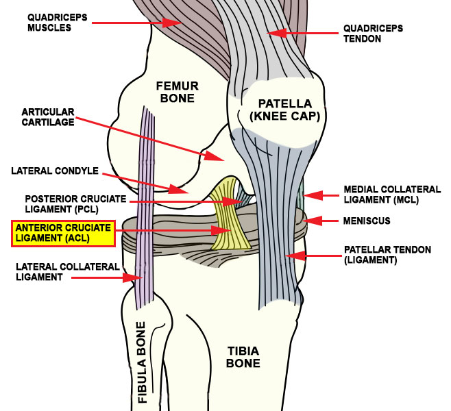
- Posterior Cruciate Ligament (PCL): Limits the anterior rolling of the femur on the tibia during extension.
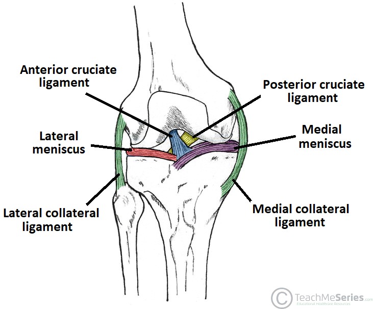
Menisci
- Medial Meniscus

- Lateral Meniscus
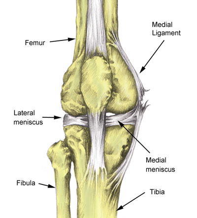
The menisci increase the articular surface area and act as a shock absorber. 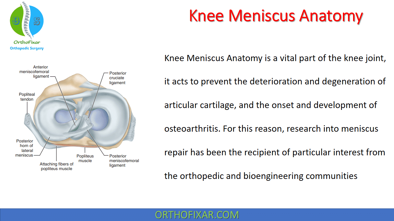
Movements of the Knee Joint
The knee joint is a hinge type of synovial joint allowing flexion and extension. It is most stable when extended. 
- Extension: By Quadriceps Femoris

- Flexion: By Hamstrings

Knee Joint Injuries
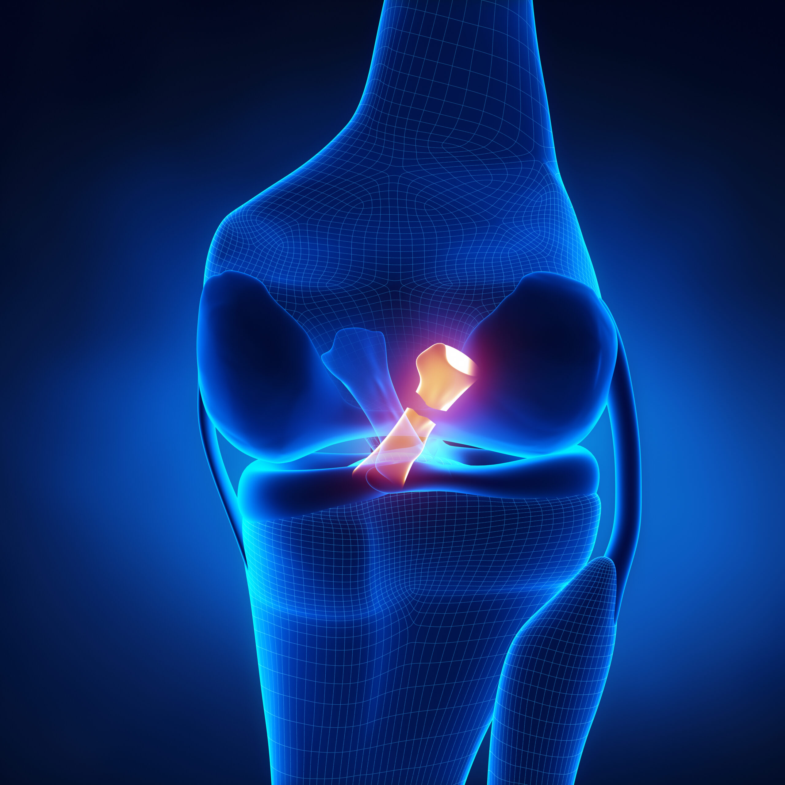
Unhappy Triad: Caused by the rupture of:
- Tibial collateral ligament

- Medial meniscus
- Anterior cruciate ligament (ACL)
- Tibial collateral ligament
ACL Rupture: Anterior drawer sign where the tibia slides anteriorly.

- PCL Rupture: Posterior drawer sign where the tibia slides posteriorly.

Compartments of the Thigh
Posterior Compartment (Flexor)
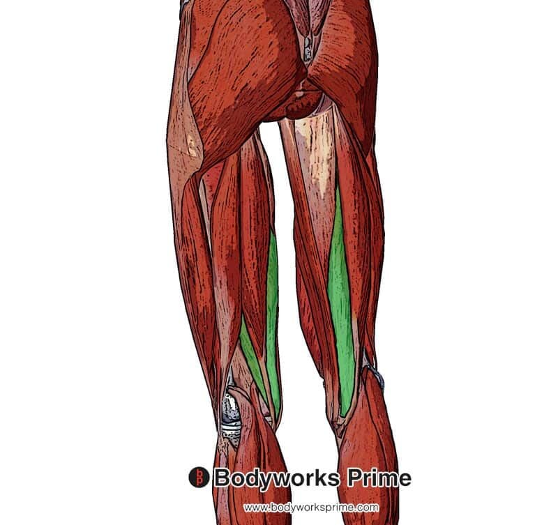
- Hamstring Muscles:
- Biceps Femoris (Long Head)
- Semitendinosus
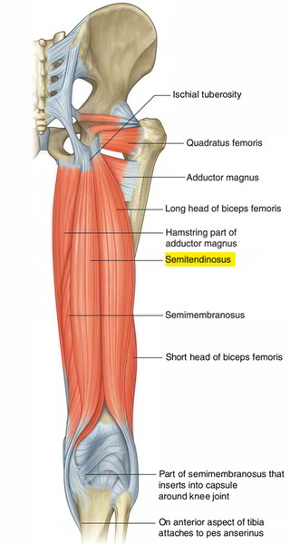
- Semimembranosus
- Biceps Femoris (Long Head)
- Innervation: Tibial nerve
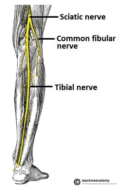
- Popliteal Vessels

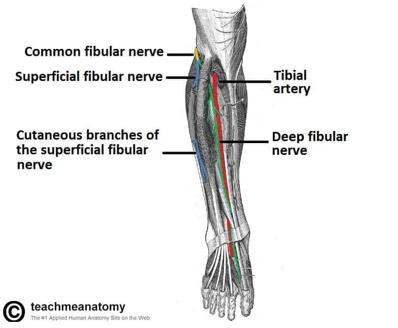
- Common Fibular Nerve

Arteries and Nerves of Thigh
- Posterior Femoral Cutaneous Nerve
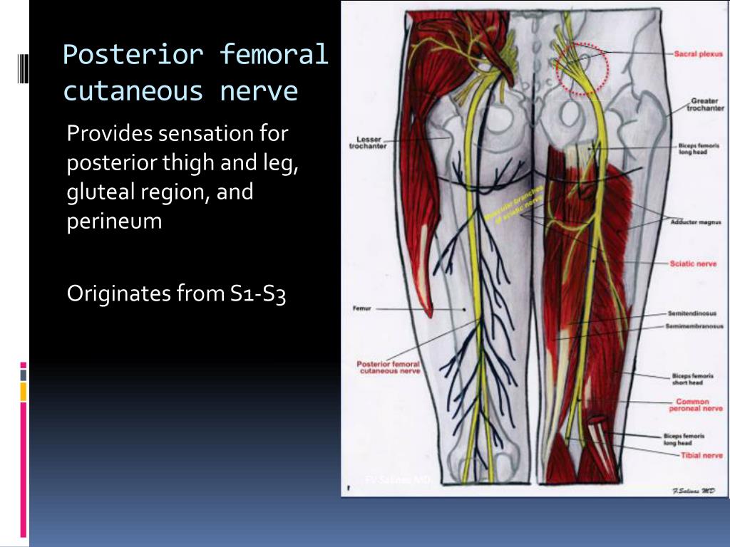
- Sciatic Nerve

- Medial Circumflex Femoral Artery
:background_color(FFFFFF):format(jpeg)/images/article/en/medial-circumflex-femoral-artery/87ujEZIAL1ccAptGRTC2PQ_4Jr73wM4q87jJ9wxWXNPg__Medial_circumflex_femoral_artery.png)
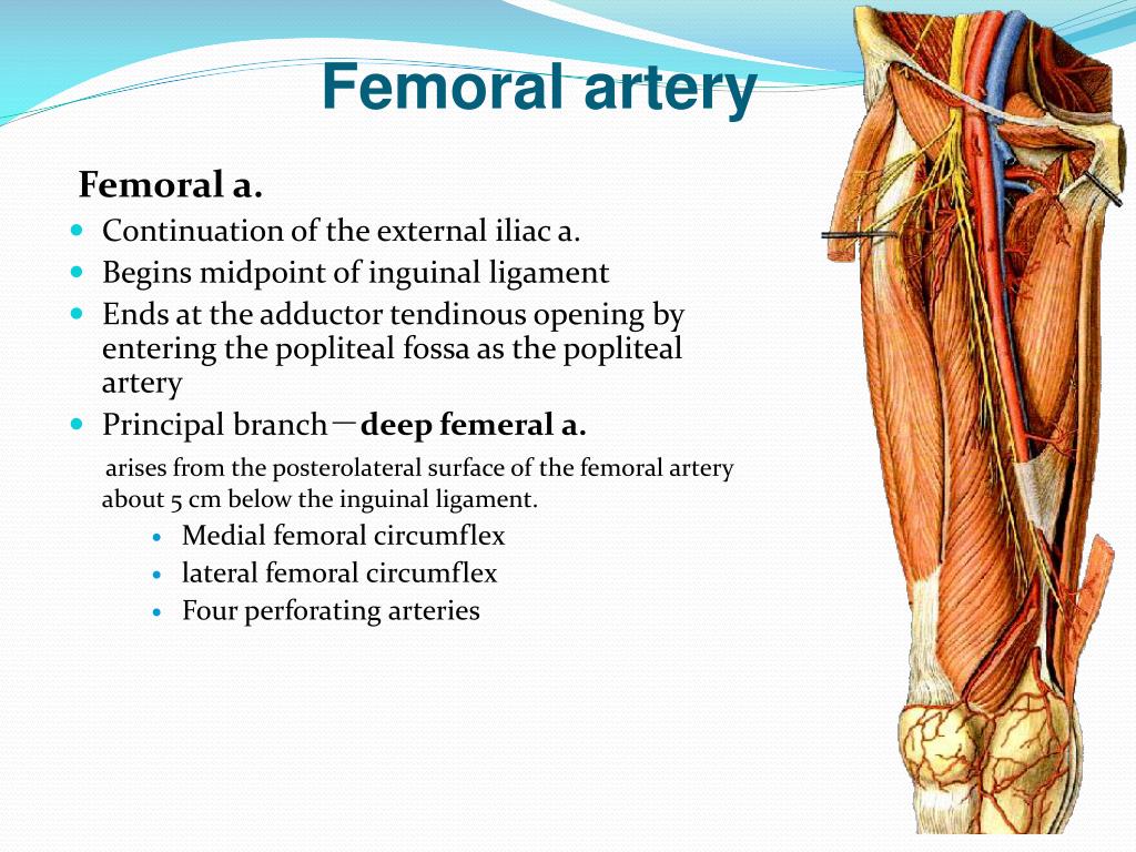
- Perforating Arteries
- Muscular Branches of Sciatic Nerve
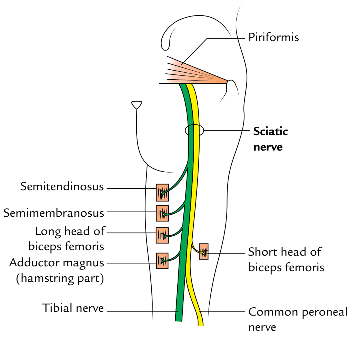
- Tibial Nerve
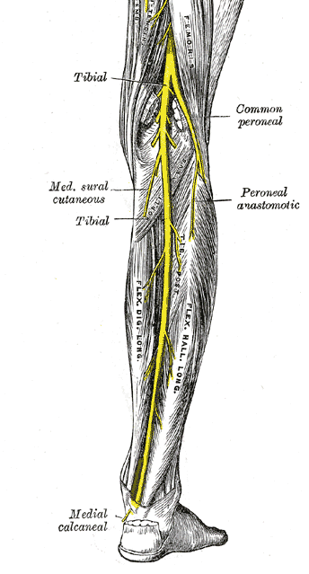
- Common Fibular Nerve
Popliteal Fossa
Contents of the Popliteal Fossa
Superficial to deep:
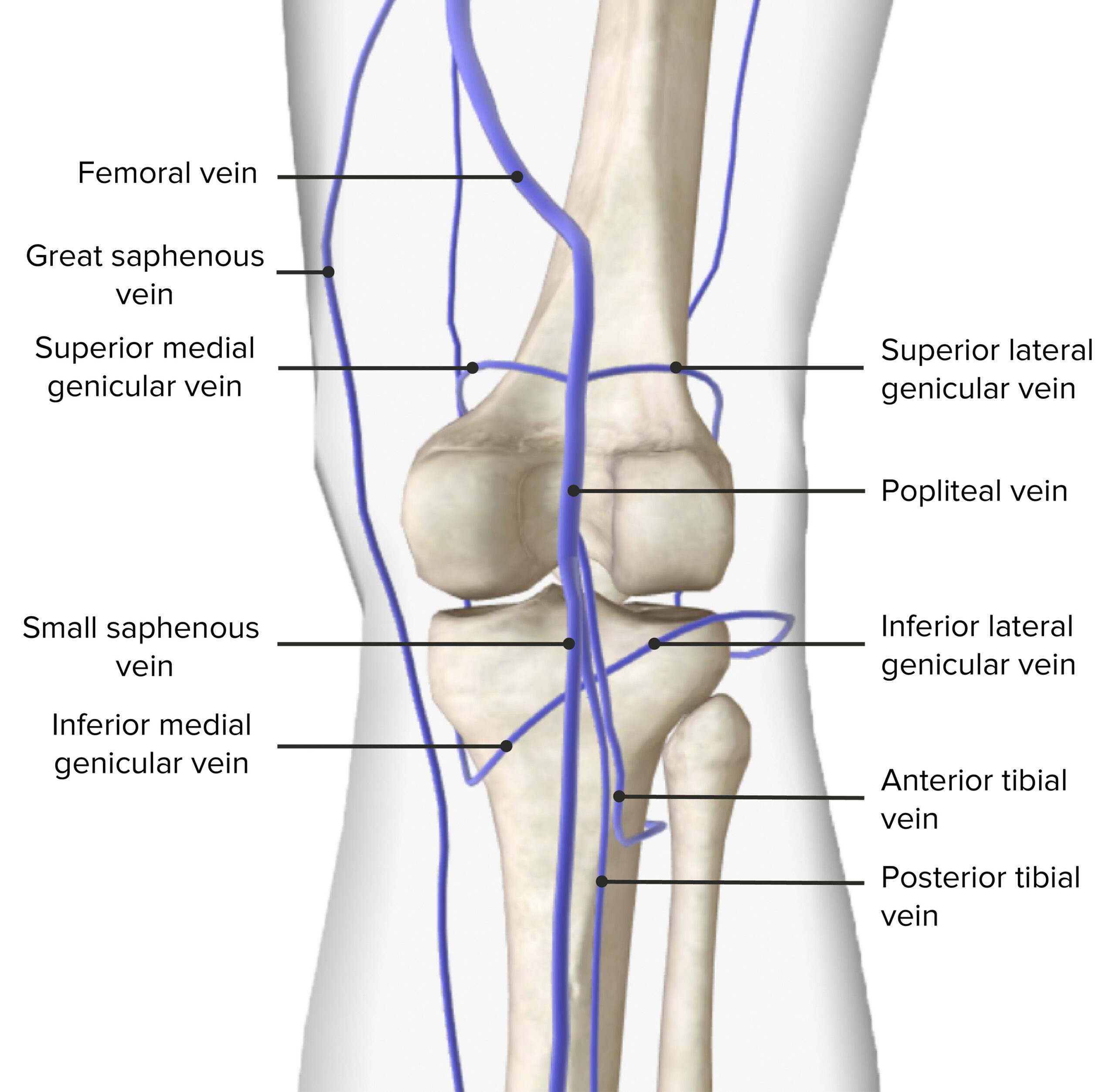
- Tibial Nerve 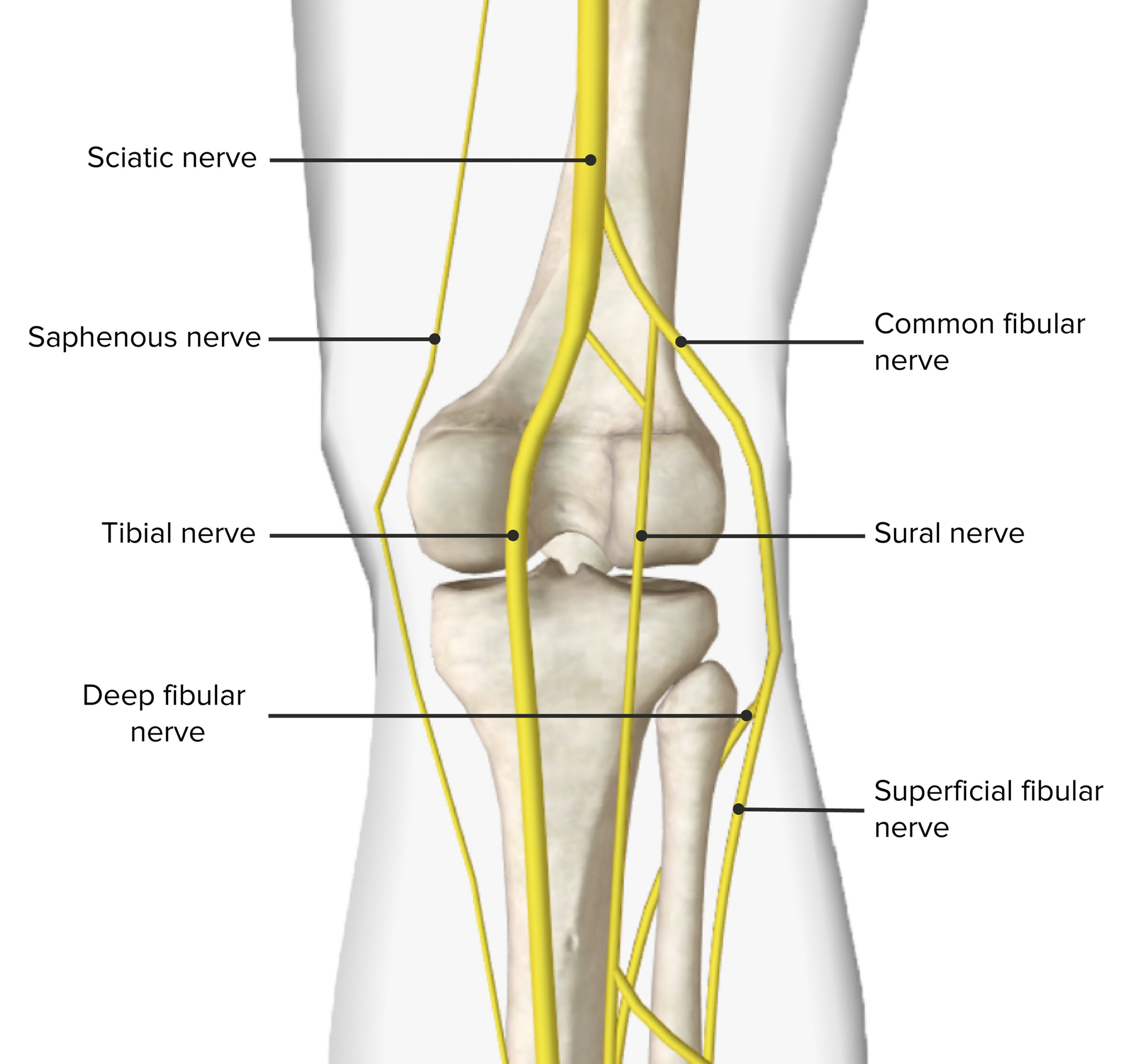 - Popliteal Vein
  - Popliteal Artery
Floor
- The popliteal surface of the femur
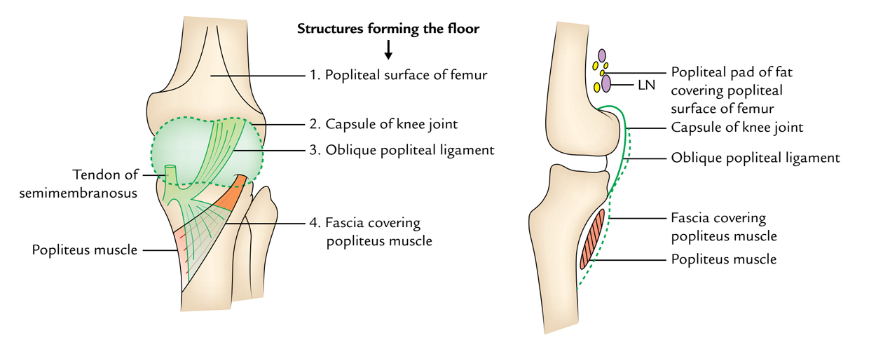
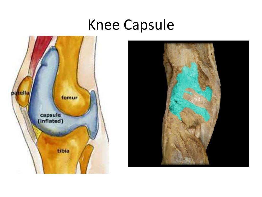
- Knee joint capsule
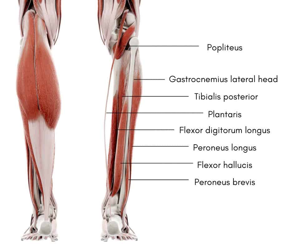
- Popliteus muscle
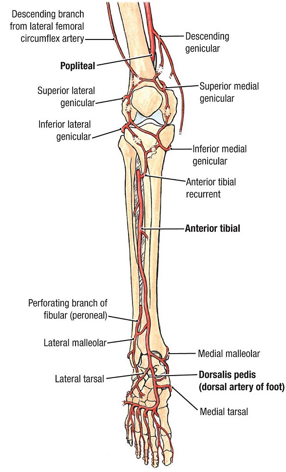
Arteries of Thigh & Knee
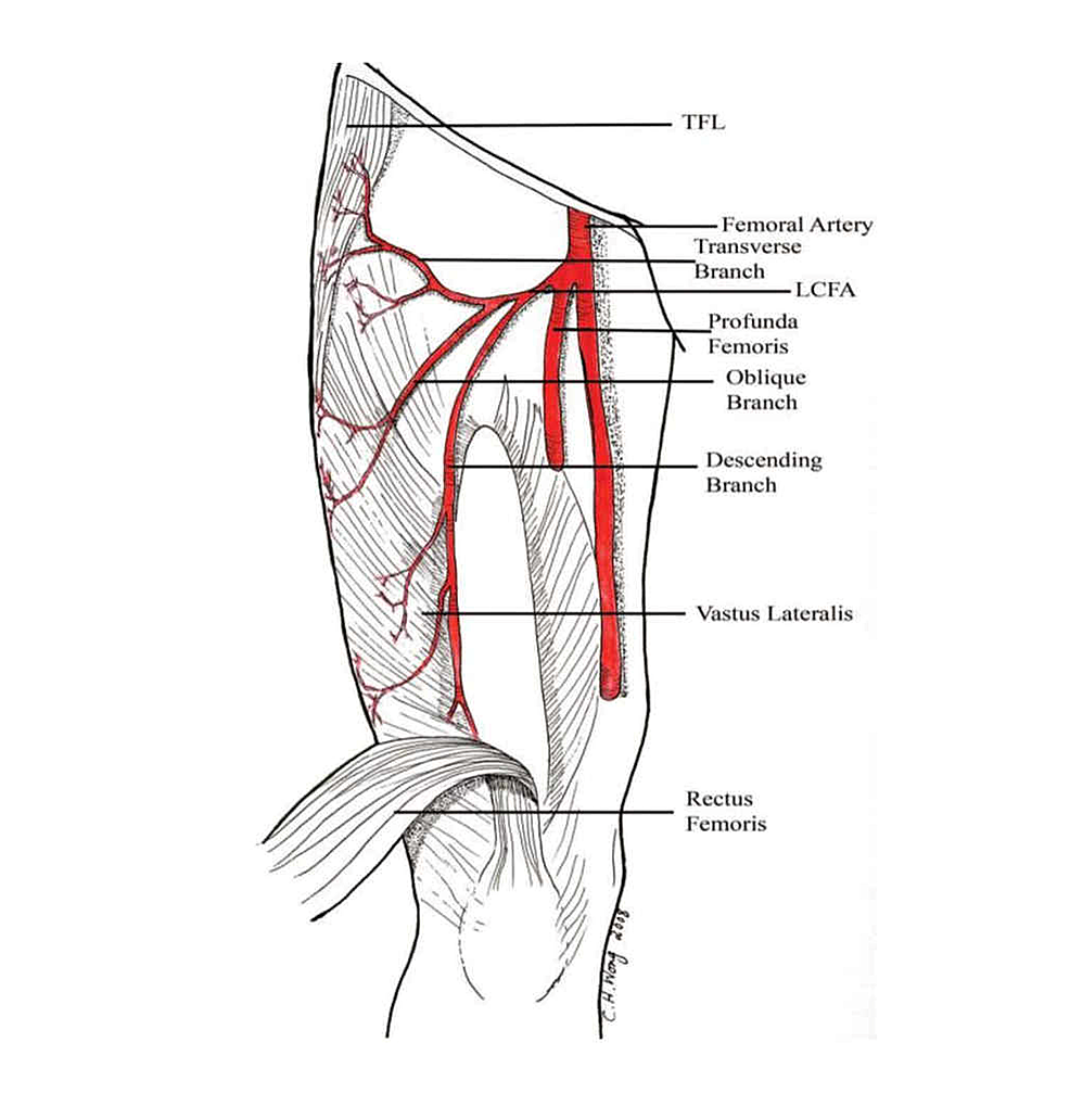
- Femoral Artery
- Popliteal Artery
- Superior Lateral Genicular Artery

- Superior Medial Genicular Artery

- Inferior Lateral Genicular Artery
:background_color(FFFFFF):format(jpeg)/images/library/7457/LD7FudenYWNGIT3Q8WbOmg_bXE1JXX9if_Arteria_inferior_lateralis_genus_1.png)
- Inferior Medial Genicular Artery

- Superior Lateral Genicular Artery
- Anterior Tibial Artery
Lymph Vessels & Nodes
- Popliteal Vein
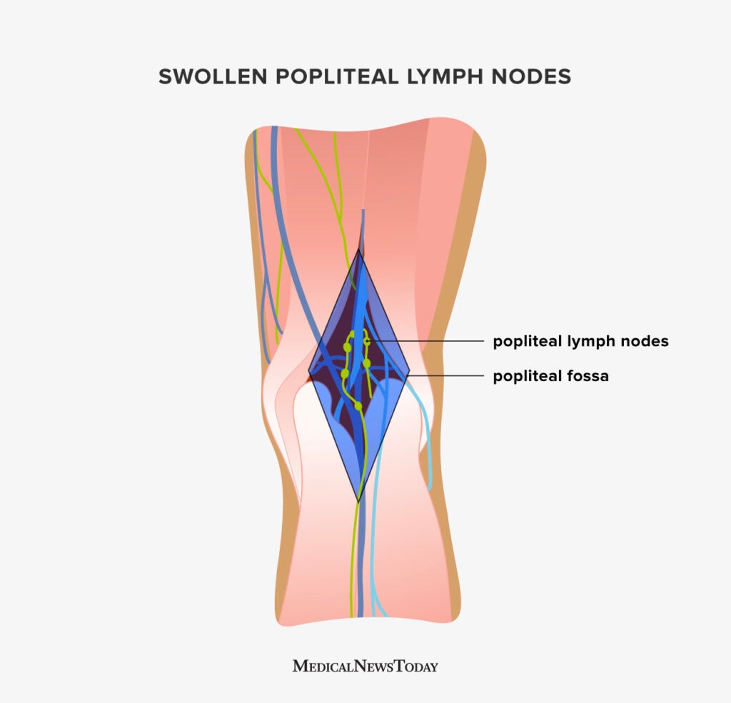
- Popliteal Lymph Node
:background_color(FFFFFF):format(jpeg)/images/library/5482/cueOOd9R1COE5GbAXKhQ_Arteria_poplitea_2.png.jpeg)
- Small Saphenous Vein

