Face, Parotid and Scalp
Face, Parotid and Scalp
Muscles of Facial Expression
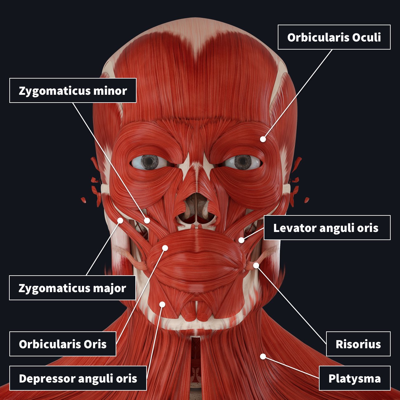
There are 21 muscles of facial expression in the face and neck, with the 12 major muscles spanning the orbital, nasal, and oral regions. All these muscles receive motor innervation from CN VII (Facial Nerve).
Major Facial Muscles
- Platysma

- Occipitofrontalis
- Frontalis (frontal belly)
:watermark(/images/watermark_only_413.png,0,0,0):watermark(/images/logo_url_sm.png,-10,-10,0):format(jpeg)/images/anatomy_term/frontalis-muscle/vxlrpq9fdi58f2MWrUDkw_frontalis_muscle.jpg)
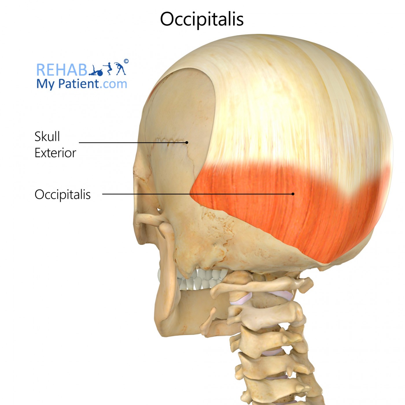
- Occipitalis (occipital belly)
- Frontalis (frontal belly)
- Orbicularis Oculi
- Orbital
- Palpebral

- Orbicularis Oris
:background_color(FFFFFF):format(jpeg)/images/article/orbicularis-oris-muscle/e1HTO3sjqPhfSGPAMu9vA_sbzEu7YwVgV3CH95mY9jhg_Orbicularis_oris_muscle_01.png)
- Mentalis
:background_color(FFFFFF):format(jpeg)/images/library/13033/XZyHj5YSlZlu070CzBopDg_Mentalis_muscle_01.png)
- Zygomaticus Major
:background_color(FFFFFF):format(jpeg)/images/library/13047/rpZWN14YWkRho1b7KJBO6w_Zygomaticus_major_muscle_01.png)
- Zygomaticus Minor
:background_color(FFFFFF):format(jpeg)/images/library/13028/GMyjaIGr4s16Njvg2oi8Yw_Zygomaticus_minor_muscle_01.png)
- Levator Labii Superioris
:background_color(FFFFFF):format(jpeg)/images/library/13024/w4XmgTQsoKjFNOeKylE39Q_Levator_labii_superioris_muscle_01.png)
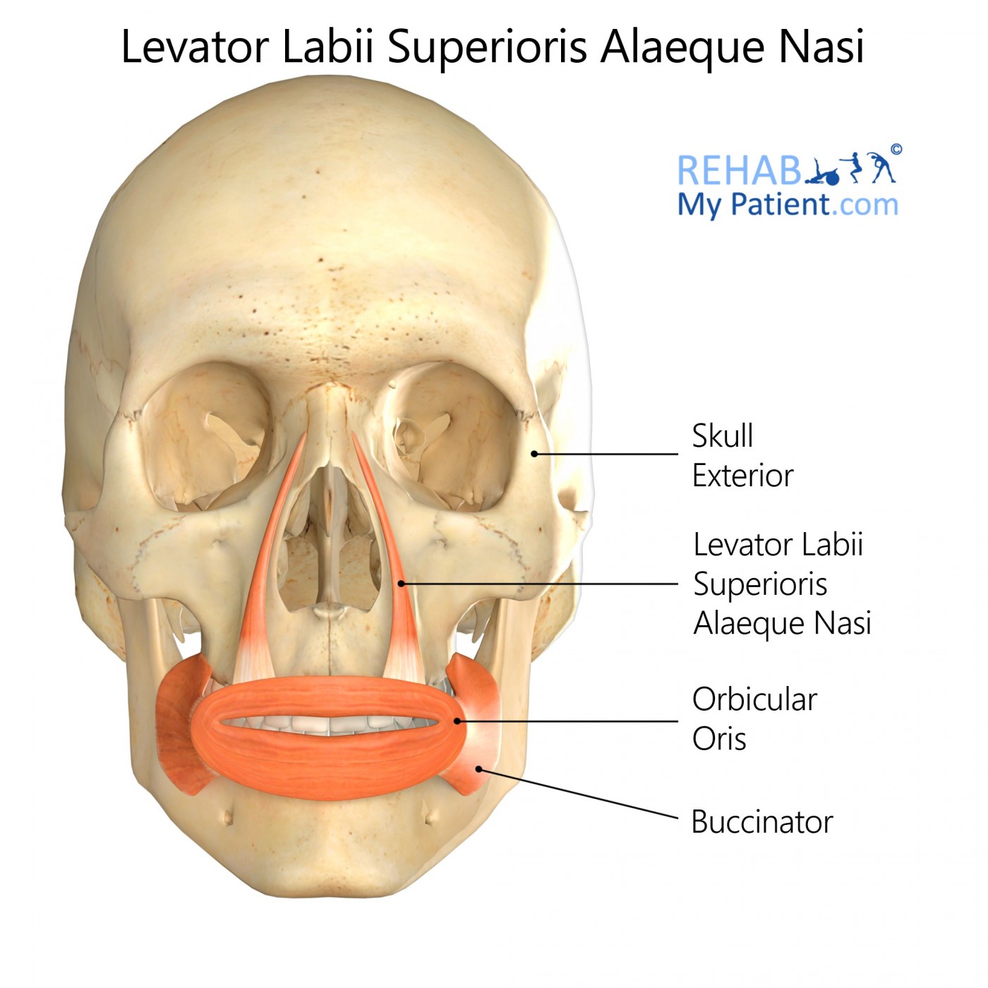
- Levator Labii Superioris Alaeque Nasi
- Levator Anguli Oris
:background_color(FFFFFF):format(jpeg)/images/library/13030/4gEpcmExWqTl8WaDXj0rGQ_Levator_anguli_oris_1.png)
- Risorius
:background_color(FFFFFF):format(jpeg)/images/library/13051/ffRKTrgzyay0Z39clZKOw_Risorius_muscle_01.png)
- Depressor Anguli Oris
:background_color(FFFFFF):format(jpeg)/images/library/13036/vzE821Hv9jhH8P7DP6kHRw_Depressor_anguli_oris_muscle_01.png)
- Depressor Labii Inferioris
:background_color(FFFFFF):format(jpeg)/images/library/13027/mV7X34SRZnOh5ZZXKl6Q_Depressor_labii_inferioris_muscle_01.png)
- Buccinator
:background_color(FFFFFF):format(jpeg)/images/library/13049/CBtkqnBzI3RakynrTNwcUQ_Buccinator_muscle_01.png)
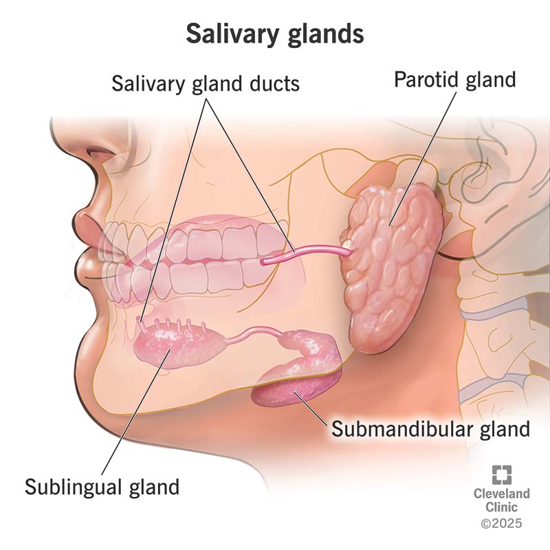
Parotid Gland
The parotid gland is located near the mandible and is associated with the parotid duct. Notably, it receives parasympathetic innervation from CN IX, with CN VII passing through it without innervating it.
Facial Nerve (CN VII)
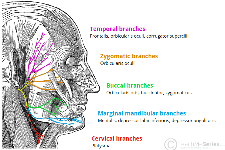
The motor branches of CN VII include:
- Temporal branches
- Zygomatic branches
- Temporofacial branch
- Posterior auricular nerve
- Buccal branches
- Cervicofacial branch
- Marginal mandibular branches
- Cervical branches
- Digastric branch
Cutaneous Distribution of CN V
CN V (Trigeminal Nerve) has three main branches, each responsible for sensory innervation to different regions of the face:
- V1: Ophthalmic nerve
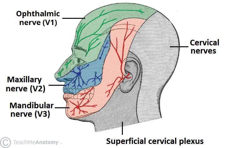
- V2: Maxillary nerve
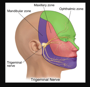
- V3: Mandibular nerve
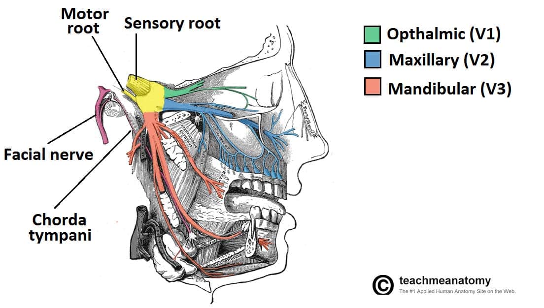
Notable Nerves
- Supra-orbital nerve

- Supratrochlear nerve

- Infratrochlear nerve
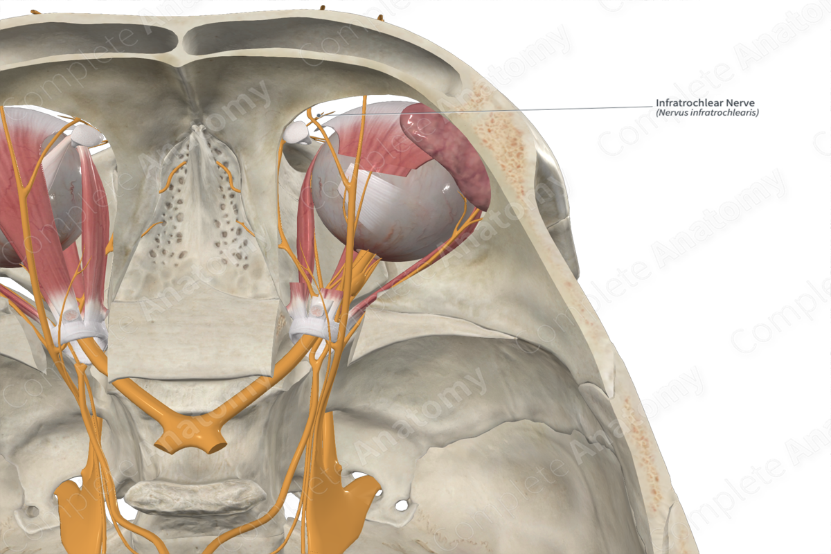
- External nasal nerve
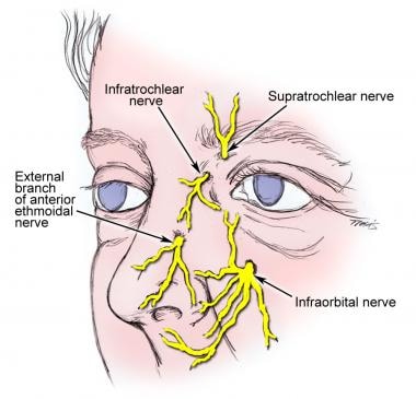
- Infra-orbital nerve

- Auriculotemporal nerve
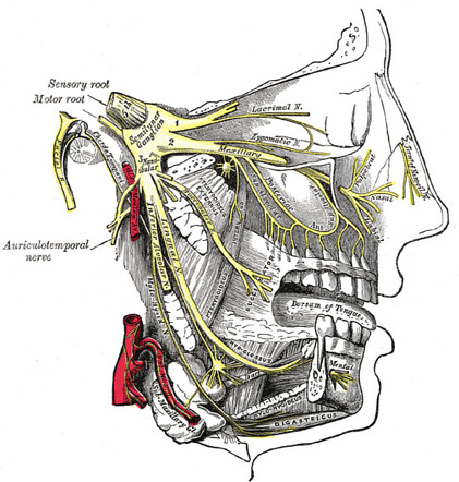
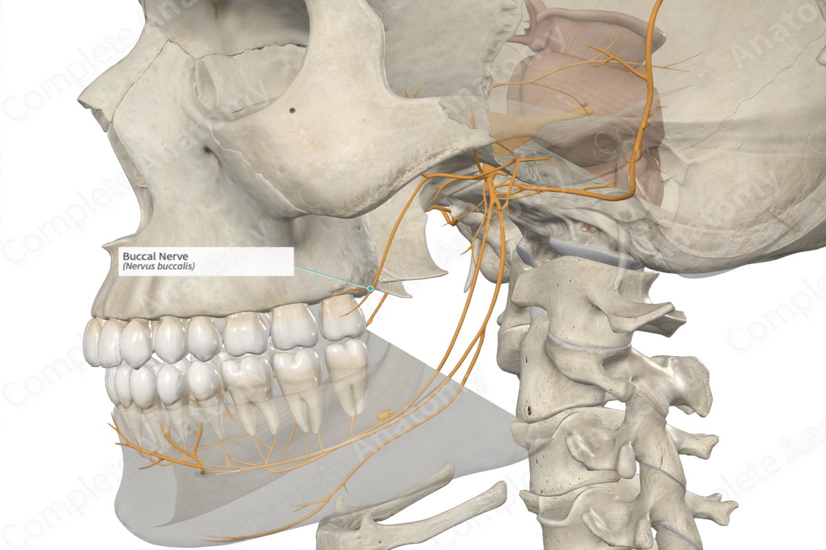
- Buccal nerve
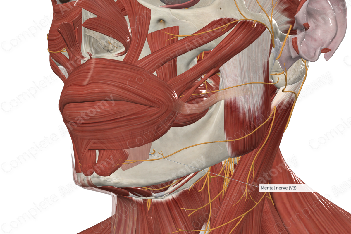
- Mental nerve
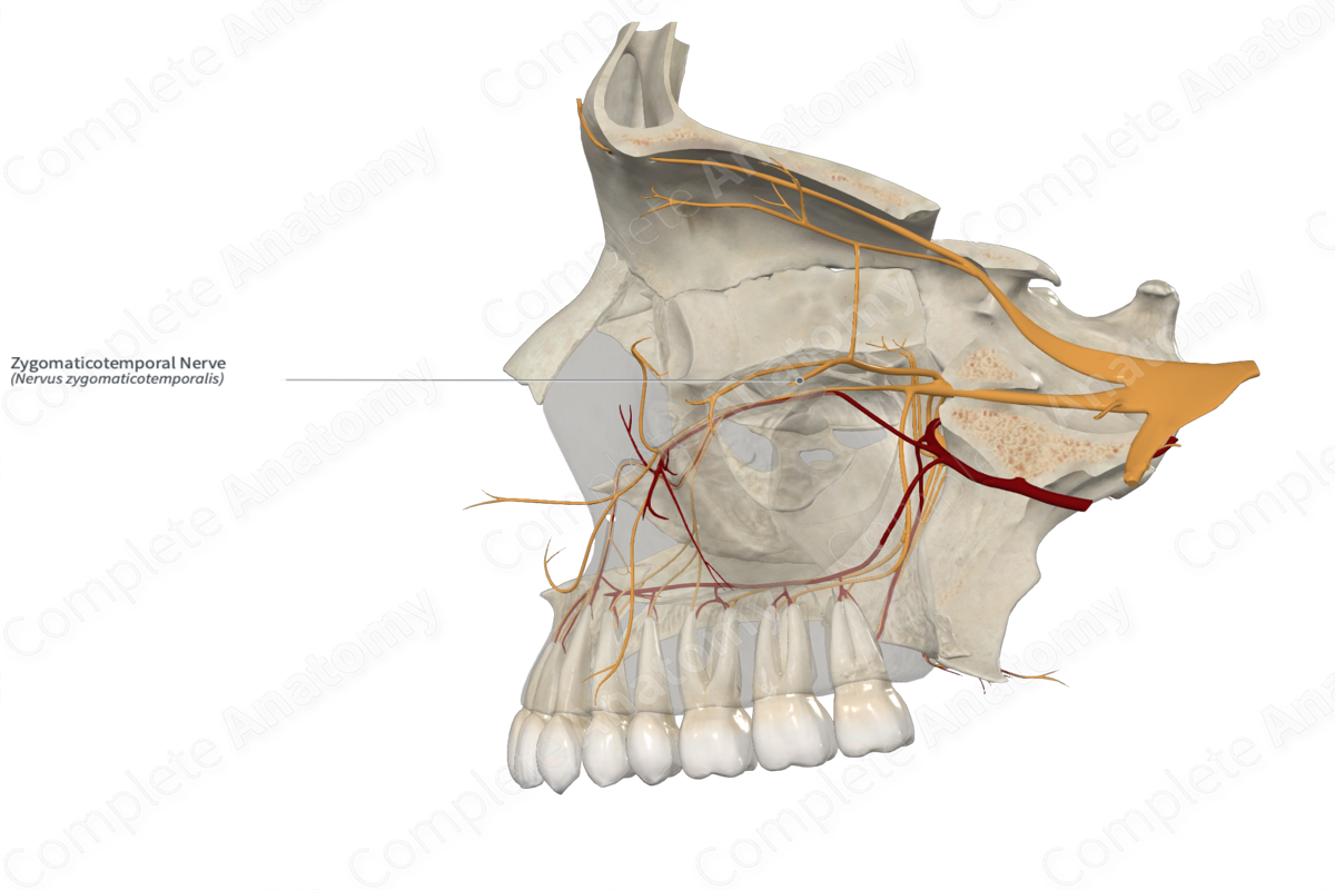
- Zygomaticotemporal nerve
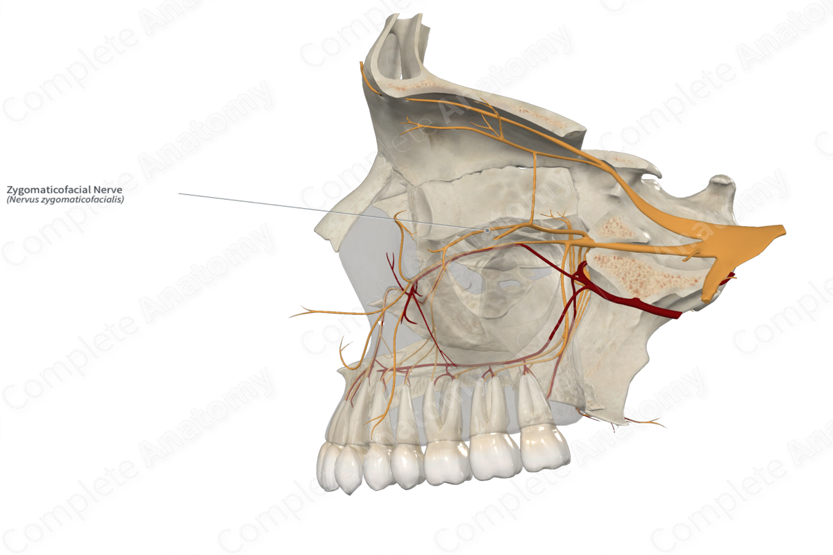
- Zygomaticofacial nerve
Pattern of Arterial Supply to the Face and Scalp
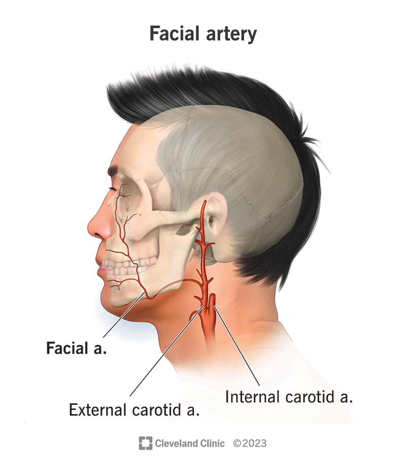
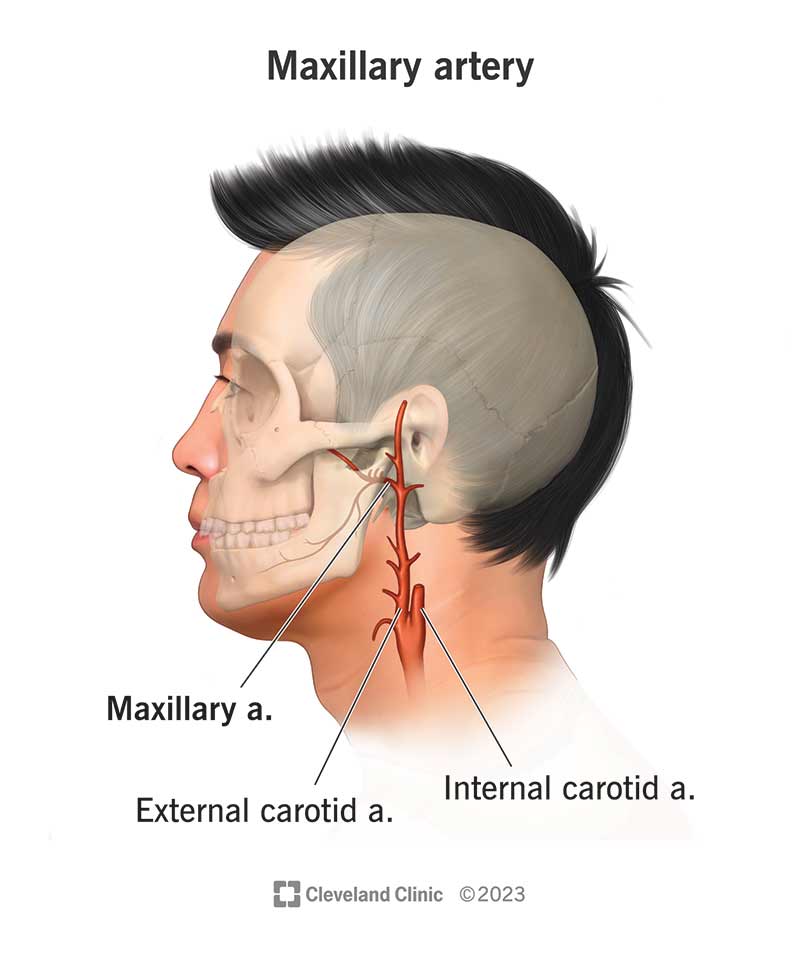
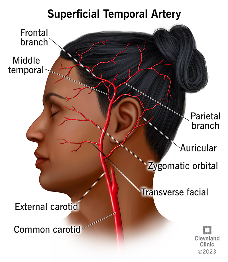
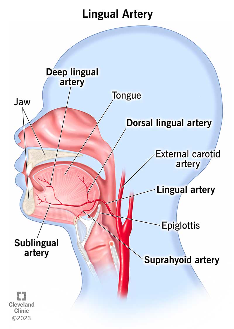
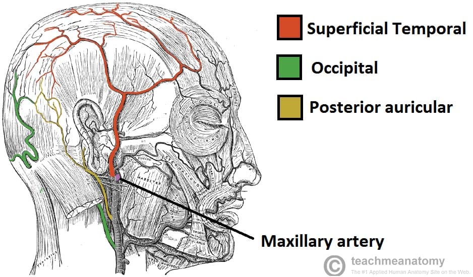
The external carotid artery and its branches supply blood to the face and scalp. Some of the major branches include:
- Facial artery
- Maxillary artery
- Superficial temporal artery
- Transverse facial artery

- Infra-orbital artery
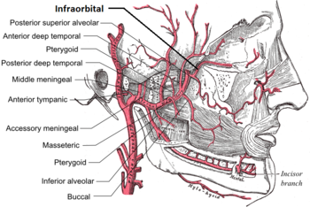
- Buccal artery

- Lingual artery
- Mental artery
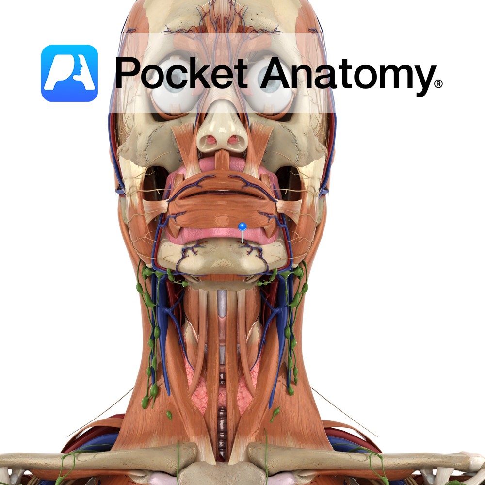
Some arteries like the supratrochlear and supraorbital arise from the internal carotid system.
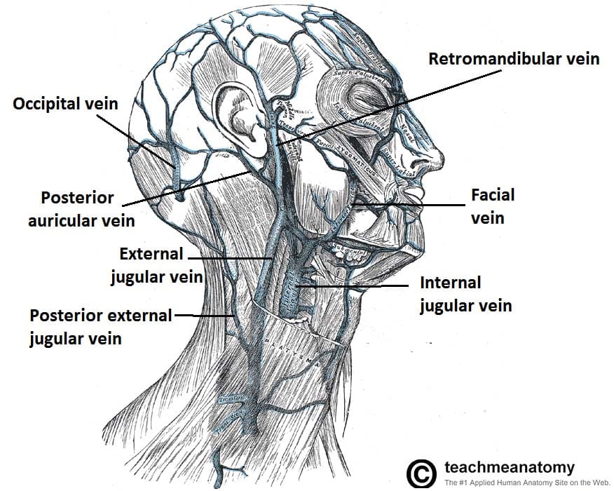
Venous Drainage of the Face
The veins of the face include the facial, angular, and ophthalmic veins, which are valveless. The facial vein drains into the internal jugular vein (IJV) while the ophthalmic veins and pterygoid plexus drain into the cavernous sinus. Anastomoses present potential danger due to possible spread of infection.
Lymphatic Drainage of the Face
The lymphatic vessels of the face primarily drain into the following nodes:
- Submental nodes
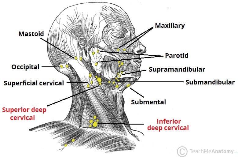
- Submandibular nodes
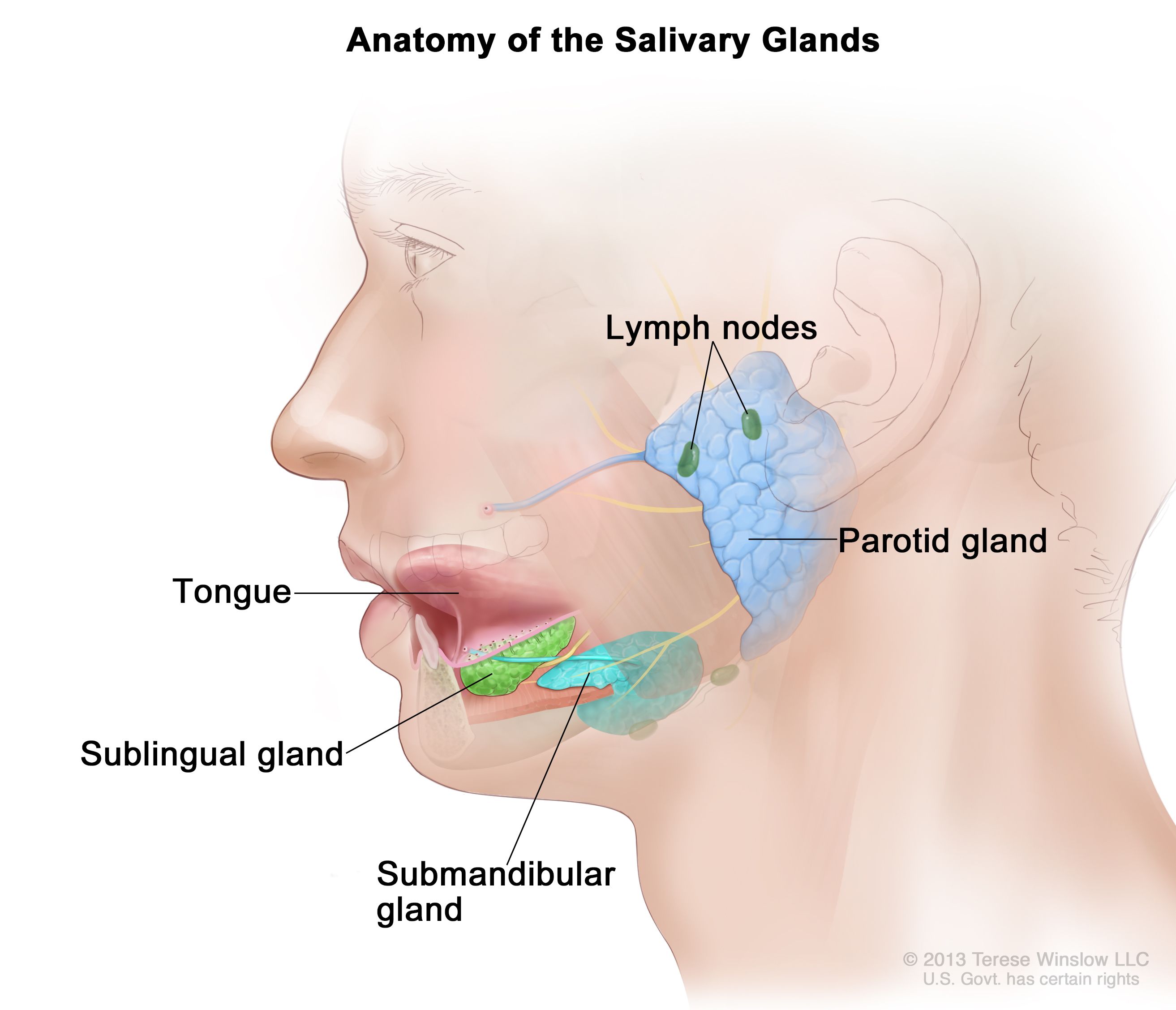
- Pre-auricular and parotid nodes

Layers of the Scalp
The scalp consists of five layers:
- Skin
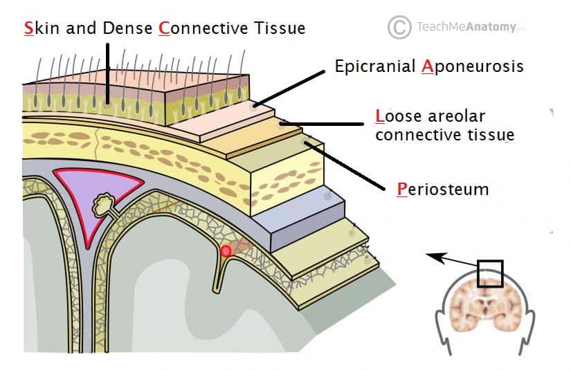
- Connective tissue

- Aponeurosis

- Loose connective tissue
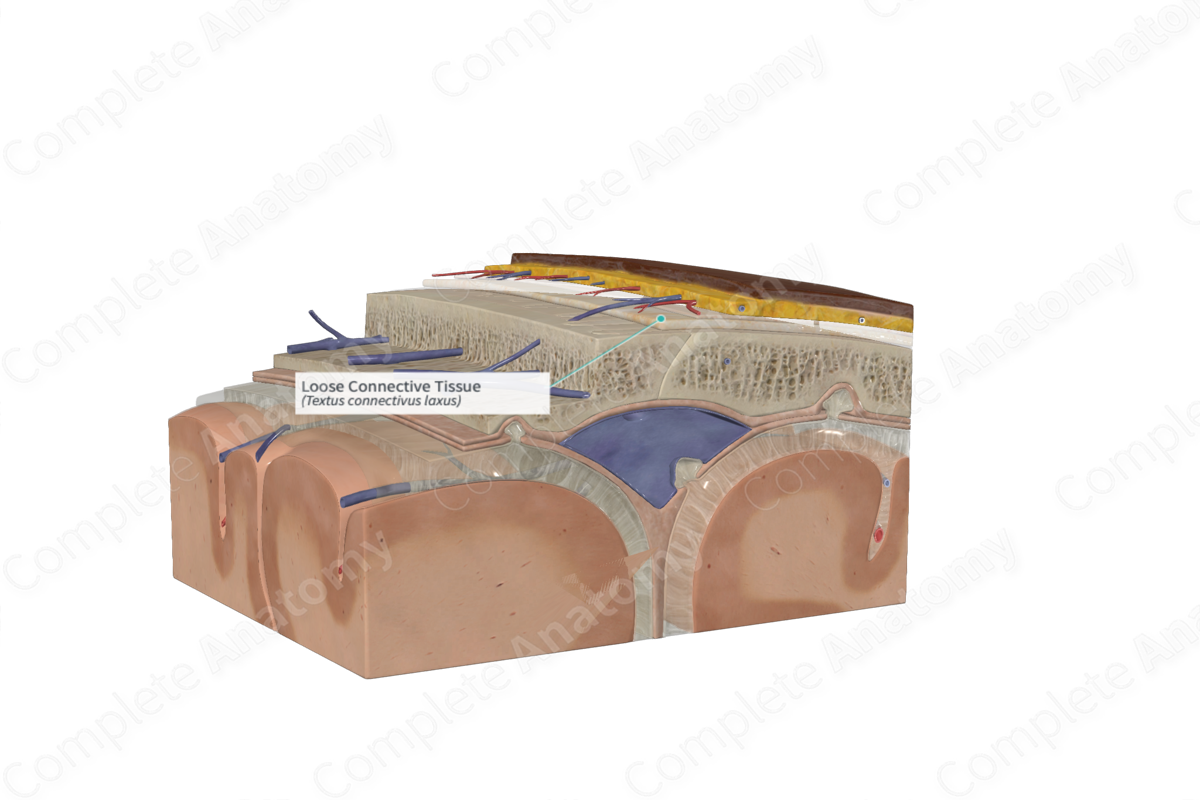
- Periosteum/Pericranium

Clinical Correlations
- Lump on the forehead

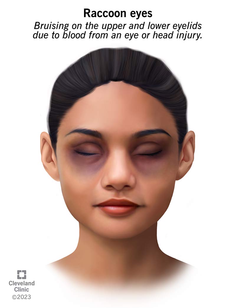
- Raccoon's eyes (periorbital ecchymosis)
- Profuse scalp bleed which is exacerbated by:
- The occipitofrontalis muscle pulling the sides of the wound apart
- Dense connective tissue holding cut vessels open
