Abdominal Wall, Inguinal Region, and Peritoneal Cavity
Anatomy of the Abdominal Wall, Inguinal Region, and Peritoneal Cavity
Surface Anatomy and Skeleton of the Abdominal Wall
- Key Landmarks:
- Xiphisternal junction
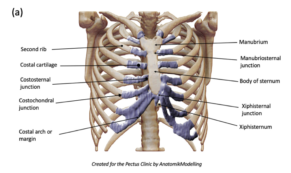
- Xiphoid process
- Costal margin

- Iliac crest
- Anterior superior iliac spine
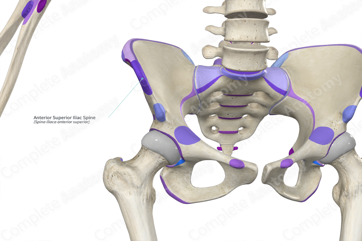
- Pubic crest

- Pubic tubercle
- Inguinal ligament
:background_color(FFFFFF):format(jpeg)/images/library/13034/pxqvPbiAM9u42mf3lX3Q_Lig._inguinale_02.png)
- *Note from an M2: remember the order of what goes under the inguinal ligament. There are many questions about the herniations and ordering here.
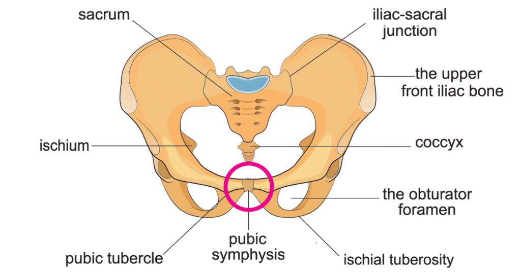
- Pubic symphysis
Regional Descriptive Terminology
Nine-Region System:
*Note from an M2: Know which organs are in which quadrants

- Hypochondriac regions (left and right):
- Right hypochondriac: Contains the right lobe of the liver, gallbladder, and part of the right kidney.
- Left hypochondriac: Contains the spleen, part of the stomach, and part of the left kidney.
- Epigastric region:
- Located in the upper middle of the abdomen.
- Contains part of the liver, part of the stomach, part of the pancreas, and the duodenum.
- Lumbar regions (left and right):
- Right lumbar: Contains part of the ascending colon and part of the right kidney.
- Left lumbar: Contains part of the descending colon and part of the left kidney.
- Umbilical region:
- The central region of the abdomen is around the navel.
- Contains parts of the small intestine, part of the transverse colon, and part of the greater omentum.
- Inguinal regions (left and right):
- Lower lateral areas of the abdomen.
- Contains parts of the small intestine and colon.
- In males, the spermatic cord passes through this region.
- Hypogastric (pubic) region:
- The lower central region of the abdomen.
- Contains the bladder (when full), parts of the small intestine, and in females, the uterus.
Remember that these regions are not strictly defined compartments; organs can overlap multiple regions. This division helps describe the location of abdominal pain or masses and perform physical examinations.
Quadrant System:
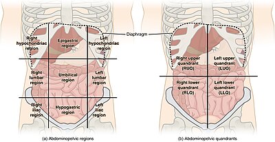
- Right Upper Quadrant (RUQ):
- Contains: Right lobe of the liver, gallbladder, right kidney, part of the pancreas, part of the duodenum, hepatic flexure of the colon
- Key points: This is where you'd typically feel pain from gallstones or liver issues
- Left Upper Quadrant (LUQ):
- Contains: Spleen, left lobe of the liver, stomach, part of the pancreas, left kidney, splenic flexure of the colon
- Key points: Pain here might indicate issues with the spleen or stomach
- Right Lower Quadrant (RLQ):
- Contains: Cecum, appendix, part of the ascending colon, right ovary, and fallopian tube (in females)
- Key points: This is where you'd typically feel pain from appendicitis
- Left Lower Quadrant (LLQ):
- Contains: Descending colon, sigmoid colon, left ovary, and fallopian tube (in females)
- Key points: Pain here might indicate issues like diverticulitis
Superficial Fascia
- Layers:
- Fatty layer (Camper’s fascia)
- Membranous layer (Scarpa’s fascia)
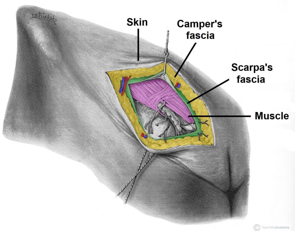
- *Note from an M2: Know the different orders of fascia around different parts of the body, including groin area.
Vessels to the Superficial Fascia of the Abdominal Wall
- Major Veins:
- Axillary vein
- Lateral thoracic vein
- Thoracoepigastric vein
- Paraumbilical veins
- Superficial epigastric vein

Cutaneous Nerves and Dermatomes
- Key Nerves:
- Subcostal nerve
- Iliohypogastric nerve
- Genitofemoral nerve

*Note from an M2: You must know the difference between these nerves and the order (from superficial to deep) that you'd find the nerves compared to the organs. Know what is deep in the genito femoral, for instance.
- Important Dermatomes:
- T6: Just inferior to the xiphoid process (not tested on this)
- T10: Skin of the umbilicus (know this)
- T12: Superior to the pubic symphysis (not tested on this)
- L1: Overlying the inguinal ligament and pubic symphysis (not tested on this)
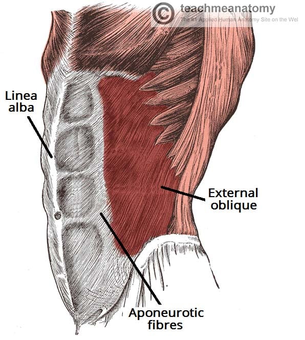
Muscles of the Anterolateral Abdominal Wall
- Layers and Functions:
- External oblique muscle
- Supports and compresses abdominal viscera
- Flexes and rotates the trunk
- Internal oblique muscle
- Transversus abdominis muscle
- External oblique muscle
Rectus Sheath
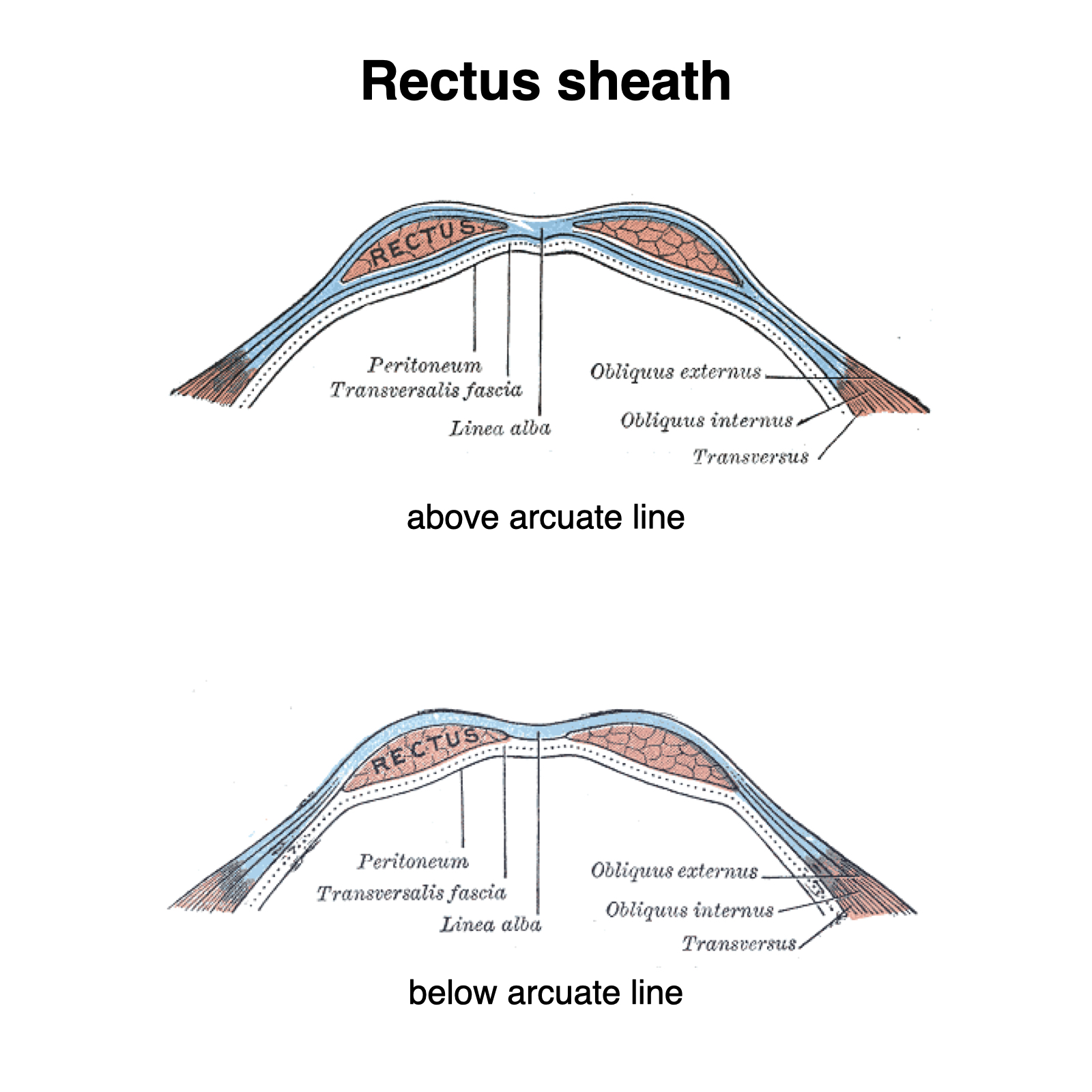
- Components:
- Rectus abdominis with tendinous intersections
- Linea alba (interlacing fibers forming the rectus sheath)
- Semilunar line
- Arcuate line (notable absence of transversus abdominis aponeurosis inferiorly)
*Note from an M2: You will be tested on these lines. Know which differences occur below and above these lines.
Inguinal Canal and Spermatic Cord
- Inguinal Hernias:
- Indirect hernias through the deep inguinal ring
- Direct hernias through the abdominal wall

Features of the Inner Surface of the Anterior Abdominal Wall
- Key Structures:
- Parietal and visceral peritoneum
- Mesenteries, omenta, ligaments, and folds of the peritoneum
*Note from an M2: You will be tested on the differences between these.
Peritoneum and Peritoneal Cavity
- Peritoneum:
- Continuous, slippery, transparent serous membrane
- Parietal Peritoneum:
- Blood, lymphatic, and somatic nerve supply identical to the wall it covers
- Sensitive to pressure, pain, heat, cold, and laceration
- Pain is well localized, except for the inferior diaphragm
- Visceral Peritoneum:
- Blood, lymphatic, and visceral nerve supply identical to the organ it covers
- Insensitive to touch, heat, cold, and laceration
- Pain is poorly localized
- Peritoneal Fluid:
- Contains water, electrolytes, leukocytes, and antibodies
- Derived from interstitial fluid in adjacent structures
- Peritoneal cavity is completely closed in males
Greater and Lesser Omentum
- Greater Omentum:
- Rich vasculature
- Provides nutrients to the peritoneal cavity

- Lesser Omentum:
- Contains important structures such as the hepatic portal vein and hepatic artery

Mesenteries
- Key Mesenteries:
- Mesentery of the small intestine
- Transverse mesocolon
- Sigmoid mesocolon
Organs
- Intraperitoneal Organs:
- Stomach, small intestine (except for parts of the duodenum), appendix, transverse colon, sigmoid colon, liver, spleen
- Retroperitoneal Organs:
- Parts of the duodenum, pancreas, ascending and descending colon, kidneys, adrenal glands

