Organization of the Deep Cervical Fascia
Fascial Organization of the Neck
*Note from an M2: I remember missing a point or two because I never bothered to learn the layers of fascia very well.
The deep cervical fascia is a complex structure in the neck that can be organized into distinct layers and compartments. Understanding these layers is essential for comprehending the anatomical relationships and potential pathways for the spread of infection.

Fascial Spaces and Layers
The deep cervical fascia is divided into several layers, each with specific contents and functions:
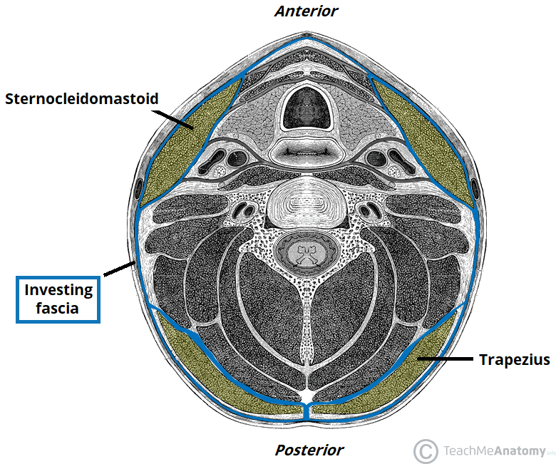
Investing Layer
- Located within: Encloses all structures of the neck except the superficial fascia
- Encloses: Trapezius and sternocleidomastoid muscles

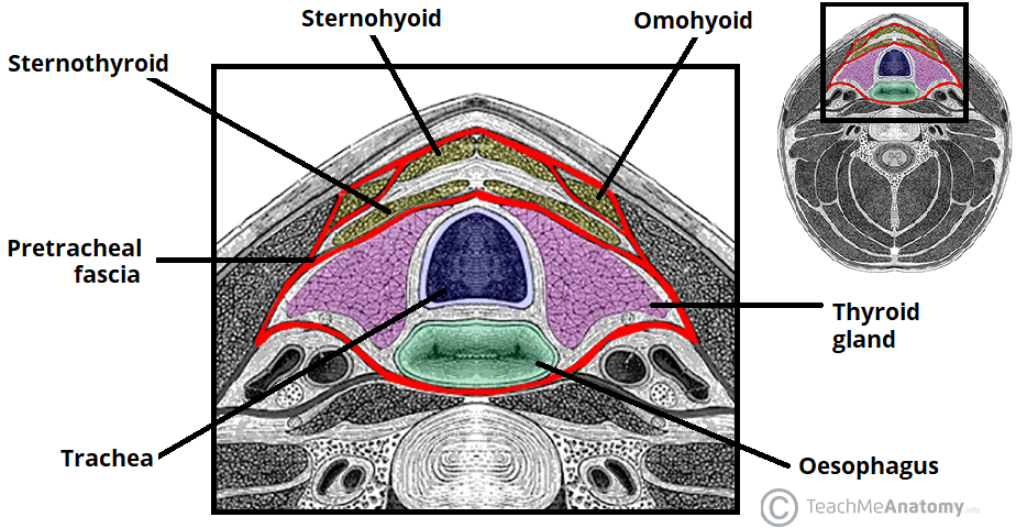
Pretracheal/Visceral Layer
- Covers: Infrahyoid muscles
- Encases: Thyroid gland, trachea, and esophagus
- Buccopharyngeal Fascia: A posterior portion of the pretracheal layer, which separates the pharynx from the prevertebral layer

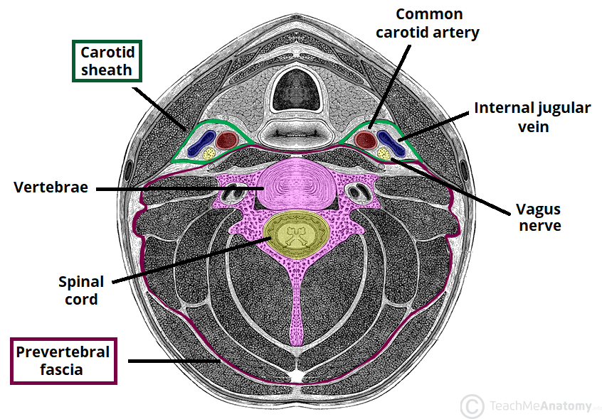
Prevertebral Layer
- Encloses: Vertebral column and associated muscles (prevertebral muscles, scalene muscles, and the deep muscles of the back)
- Fascial Space: The potential space within this layer is referred to as the 'Third Space' or 'Danger Space', which allows for the spread of infections from the neck to the thorax

Carotid Sheath
- Contains: Common carotid artery, internal jugular vein, and vagus nerve
- Provides a channel: For neurovascular structures from the base of the skull to the mediastinum
Fascial Compartments
The neck is divided into distinct fascial compartments by these fascial layers:
- Investing Layer: Encircles the entire neck just deep to the superficial fascia
- Pretracheal Space: Located within the pretracheal layer
- Retropharyngeal Space: Found between the buccopharyngeal fascia and the prevertebral fascia
- Third Space (Danger Area): A potential space located within the prevertebral layer

Clinical Correlations
Understanding the organization of the deep cervical fascia is essential for several clinical reasons:
- Compartmentalization: Fascia helps limit the spread of infections within the neck
- Support and Movement: It supports the visceral structures and facilitates movement
- Surgical Consideration: Natural cleavage planes formed by the fascial layers are important in surgical procedures
- Communication with the Thorax: Some spaces allow for potential spread of infections to the thorax (mediastinum)

Key Anatomy Terms
- Manubrium of sternum: Lower boundary of the pretracheal space
- Thyroid and Esophagus: Enveloped by the pretracheal/visceral fascia
- Trachea: Encased by the pretracheal fascia
- Sternocleidomastoid Muscle: Within the investing layer
- Internal Jugular Vein, Common Carotid Artery, Vagus Nerve: Contained within the carotid sheath