Axilla: Boundary & Contents
Boundary of Axilla
Notes from an M2:
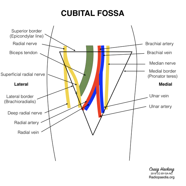
I missed several points throughout anatomy because I did not nail down the walls of the axilla. Know which organs go in which spaces, and be familiar with the barriers. You'll get about 3 points from knowing the brachial plexus diagram. Lymphatics are lower yield, just know the basics. You will be tested repeatedly on the cubital fossa. Remember
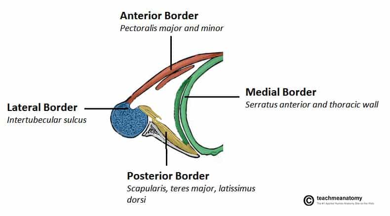
Walls of the Axilla
- Anterior Wall:
- Formed by the lateral part of the pectoralis major, pectoralis minor muscles, and clavipectoral fascia.
- The inferior most part of the anterior wall is the anterior axillary fold.
- Posterior Wall:
- Comprised of the subscapularis muscle, the distal part of the latissimus dorsi, and the distal part of teres major muscle.
- The inferior most part of the posterior wall is the posterior axillary fold.
- Medial Wall:
- Formed by the upper thoracic wall and serratus anterior muscle.
- Lateral Wall:
- Narrow and formed by the intertubercular sulcus of the humerus.

Other Boundaries
- Apex (Inlet):
- Continuous superiorly with the neck.
- Margins formed by the lateral border of the 1st rib (medially), the posterior surface of the clavicle (anteriorly), and the superior border of the scapula (posteriorly).
- Base (Floor):
- Formed by the skin of the armpit.
- Opens laterally into the arm.

Contents of the Axilla
Neurovascular Structures
- Axillary Artery:
- Branches include the superior thoracic artery, thoraco-acromial artery, lateral thoracic artery, subscapular artery, anterior circumflex humeral artery, and posterior circumflex humeral artery.
- Axillary Vein:
- Tributaries include the cephalic vein, basilic vein, and paired brachial veins, with branches corresponding to the axillary artery.
- Brachial Plexus:
- Includes roots (C5-T1), trunks, divisions, cords, and terminal branches such as the musculocutaneous nerve, median nerve, ulnar nerve, radial nerve, and axillary nerve.
Lymphatic Structures
- Lymph Nodes:
- Arranged in 5 groups (apical, central, subscapular, humeral, and pectoral), named according to their location in the axilla.
- Responsible for receiving 75% of the drainage from the breast.
- Axillary Process of the Breast:
- Extends into the axilla for additional lymphatic drainage.
Other Notable Structures
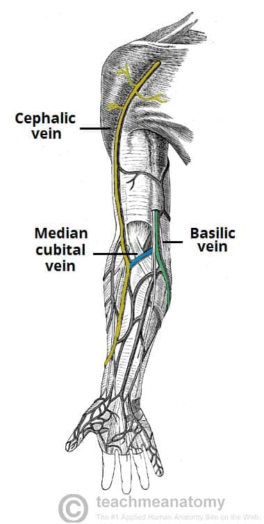
- Superficial Veins:
- Include the cephalic, median cubital (frequently used for venipuncture), and basilic veins.
- Muscle contractions help push venous blood toward the heart, while one-way valves prevent backflow.


Here's a song to help you study:
