Pharynx and Cervical Esophagus
Pharynx and Cervical Esophagus
Lecture Objectives
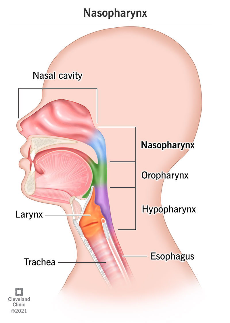
Divisions of the Pharynx
*Note from an M2: For context, these are the triangles that matter. They love testing boundaries. Know which muscles comprise which boundaries.

Nasopharynx
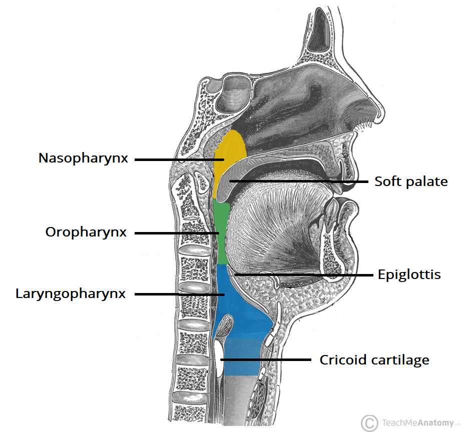
The nasopharynx is the upper part of the pharynx, connecting with the nasal cavity above the soft palate. It is bounded:
- Superiorly: By the sphenoid's body and the occipital bone's basilar part.
- Inferiorly: By the soft palate.
- Anteriorly: By the choanae (posterior nasal apertures).
- Posteriorly: By the pharyngeal tonsil and the pharyngeal recess.
Oropharynx
The oropharynx is situated behind the oral cavity. Its boundaries are:
- Superiorly: By the soft palate.
- Inferiorly: By the upper border of the epiglottis.
- Anteriorly: By the anterior pillars of the fauces.
- Posteriorly: By the bodies of the C2 and C3 vertebrae.
Laryngopharynx
The laryngopharynx is the lower part of the pharynx, lying behind the larynx and continuous with the esophagus. Boundaries include:
- Superiorly: By the upper border of the epiglottis.
- Inferiorly: By the lower border of the cricoid cartilage where it becomes continuous with the esophagus.
- Anteriorly: By the inlet of the larynx and the posterior surface of the larynx.
- Posteriorly: By the bodies of the C3 to C6 vertebrae.
External Constrictor Muscles
These muscles constrict the pharyngeal diameter during swallowing:
- Superior constrictor: Arises from the pterygoid hamulus, pterygomandibular raphe, and mandible.
- Middle constrictor: Arises from the stylohyoid ligament and the hyoid bone.
- Inferior constrictor: Arises from the oblique line of the thyroid cartilage and the cricoid cartilage.
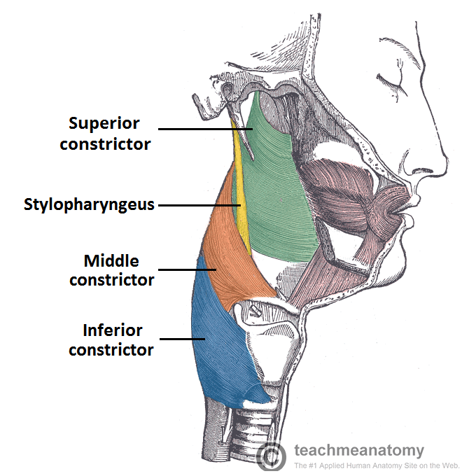
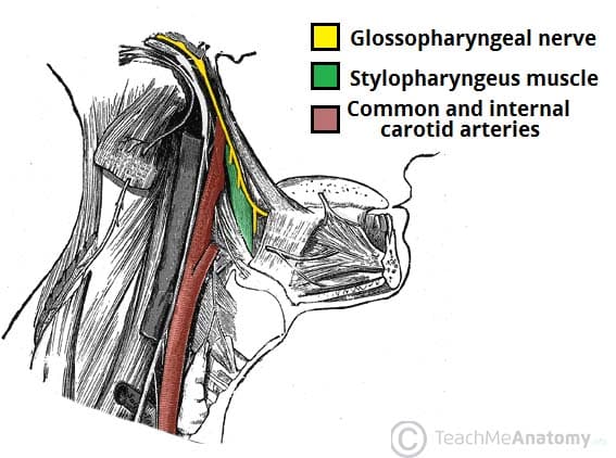
These muscles meet posteriorly at the midline raphe and are innervated by CN X, except the stylopharyngeus which is innervated by CN IX.
Longitudinal Muscles
These muscles elevate the pharynx and larynx during swallowing and speaking:
- Stylopharyngeus: Arises from the styloid process and is innervated by CN IX.
- Palatopharyngeus: Arises from the hard palate and palatine aponeurosis; innervated by CN X.
- Salpingopharyngeus: Arises from the cartilaginous part of the pharyngotympanic tube and is innervated by CN X.

Neurovascular Structures
The pharyngeal plexus provides innervation and consists of motor fibers from CN X, sensory fibers from CN IX and CN X, and vasomotor fibers (SNS) from the superior cervical ganglion.
- Nasopharynx: Sensory from CN V2 (pharyngeal branch).
- Oropharynx: Sensory from CN IX.
- Laryngopharynx: Sensory from CN X.
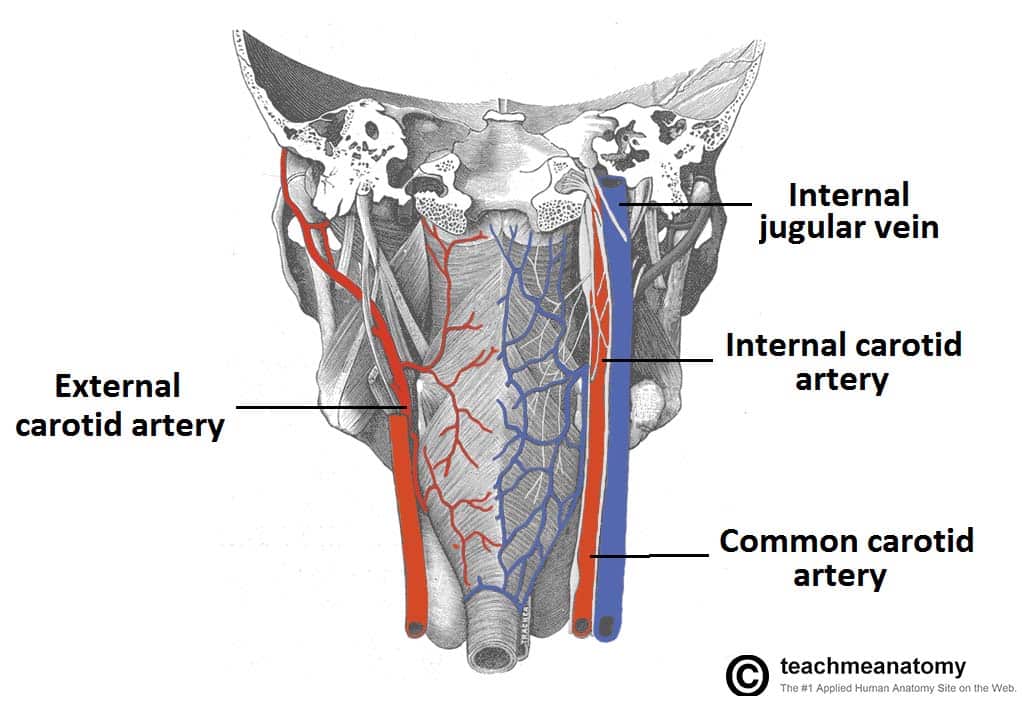
The vessels supplying the pharynx include branches from the facial artery, maxillary artery, ascending pharyngeal artery, and the inferior thyroid artery.
Cervical Esophagus
The cervical esophagus extends from the lower border of the cricoid cartilage to the thoracic inlet. It passes through the thoracic course of the esophagus within the superior and posterior mediastina.
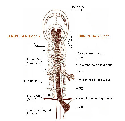
Identifying Structures
- Cervical Sympathetic Trunk: Located posterior to the carotid sheath and anterior to the prevertebral fascia.
- Vagus Nerve (CN X): Found within the carotid sheath along with the common carotid artery and internal jugular vein.
Muscular Layers of Pharynx
- Buccopharyngeal fascia on outside of the pharynx.
- Pharyngobasilar fascia lines the inside.
- Circular and longitudinal muscles meet posteriorly at the midline raphe and are mainly innervated by CN X, except stylopharyngeus (CN IX).

Gag Reflex
The gag reflex involves sensory input from CN IX and motor output from CN X.
Mucosal Coverings
- Nasopharynx: Nasopharyngeal tonsil (adenoids), torus tubarius, orifice of pharyngotympanic tube, salpingopharyngeal fold.
- Oropharynx: Palatopharyngeal pillar, palatoglossal pillar, palatine tonsil.
- Laryngopharynx: Epiglottis, vestibular fold, vocal fold, cricoid cartilage.

