Principles of Ultrasound
1. Basic Principles of Ultrasound

- Frequency refers to the number of cycles of a sound wave that occur in one second, measured in Hertz (Hz).
- Wavelength is the distance that a sound wave travels in one cycle. It is inversely related to the frequency: as the frequency increases, the wavelength decreases.
Understanding the relationship between frequency and wavelength is crucial because it affects the resolution and depth of penetration of the ultrasound waves. Higher frequencies provide better resolution but less penetration, making them suitable for imaging superficial structures. Lower frequencies penetrate deeper but with lower resolution, making them ideal for imaging deeper structures.
2. Ultrasound Transducers
Transducers are the devices that convert electrical signals into ultrasound waves and then back into electrical signals for image creation. Different transducers are used depending on the clinical application:
Linear Probe: Used for imaging superficial structures like tendons and superficial vessels. It operates at a high frequency, providing detailed images but with limited penetration.
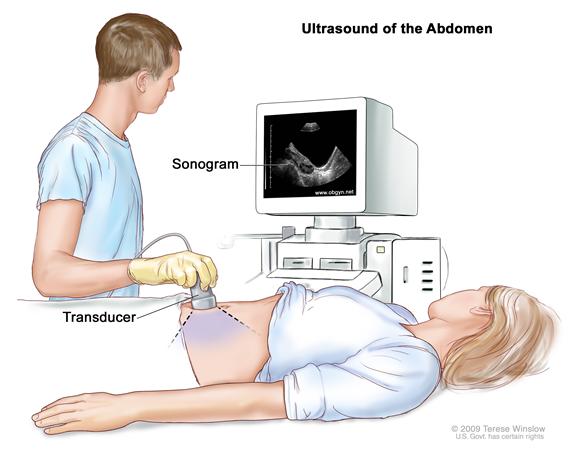
Curved Linear Probe: This is often used for abdominal imaging. It operates at a lower frequency compared to the linear probe, allowing deeper penetration but with reduced resolution.
Endocavitary Probes: These include vaginal and rectal probes used for imaging internal organs. They typically use high-frequency waves for detailed imaging of the structures within the body cavity.
3. Clinical Applications
 Ultrasound is widely used in various clinical settings due to its non-invasive nature and ability to provide real-time imaging.
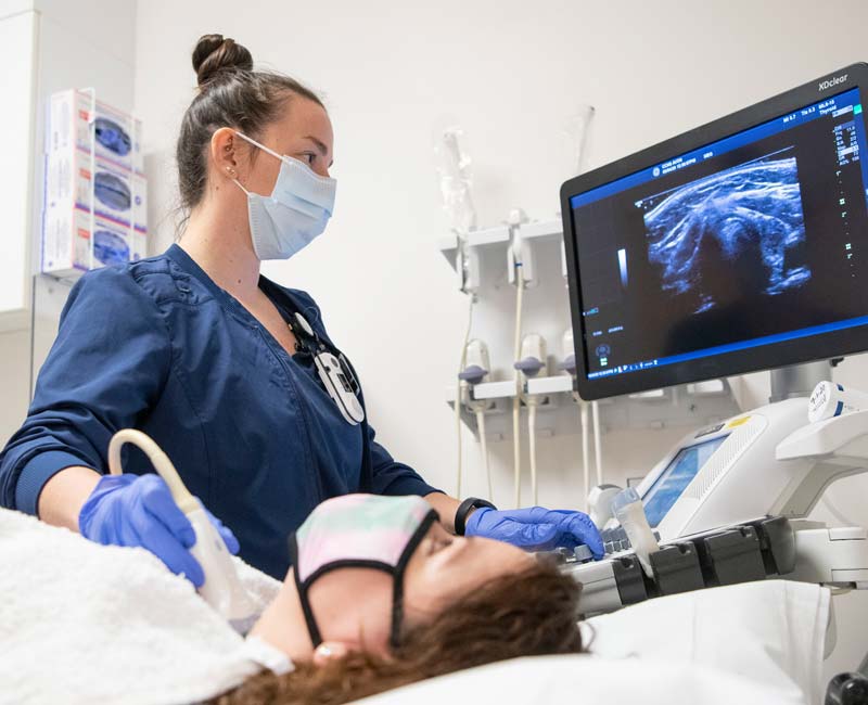
- Musculoskeletal Imaging: High-frequency linear probes are used to visualize tendons, muscles, and ligaments.
- Abdominal Imaging: Curved linear probes help in visualizing organs such as the liver, gallbladder, and kidneys.
- Obstetrics and Gynecology: Endocavitary probes are used for detailed imaging of reproductive organs and during pregnancy to monitor fetal development.
4. Key Takeaways
- The frequency and wavelength of ultrasound waves are inversely related, affecting the resolution and depth of imaging.
- Different transducers are selected based on the required resolution and depth of the target tissue.
- Ultrasound is versatile, with applications ranging from musculoskeletal assessments to internal organ imaging.